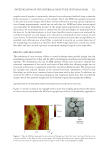
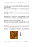
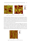
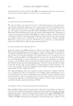
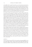
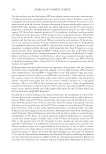
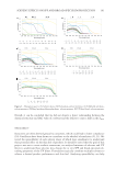
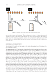
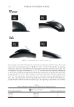
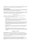
JOURNAL OF COSMETIC SCIENCE 122 the AFM image are the multiple layers of lamellar cuticle cells—roughly seven layers— surrounding the cortex of hair. A m agnifi ed view of the cuticle cells is provided in Figure 2. Within the cuticle cells, density variations are observed in the image because of the different lamellar structures within each cuticle cell. Starting from the outside and going inward toward the cortex, each individual cuticle cell, or layer, consists of the following components with thickness indicated: A layer (110 nm), exocuticle (100–300 nm), endocuticle (50–300 nm), and inner layer (10–40 nm) (42). Eac h cuticle cell is connected together by the cell membrane complex, which comprised a lipid layer on each side of the delta layer, known as the lower and upper layers, which are 2.5–4.0 nm thick. The delta layer is thought to serve as the intercellular cement between adjacent cuticle cells and believed to be composed mostly of polysaccharide. Overall, the cell membrane complex is roughly 25–28 nm thick (42). Some apparent features in the AFM micrograph of the cuticle cells are the various lamellar structures, which are out- lined in the fi gure. Sev eral key features are evident when comparing the A layer, exocuticle, and endocuticle. Not surprisingly, the A layer, which is the mostly densely crosslinked sublamina struc- ture in terms of cystine and isodipeptide crosslinks, is the least granular layer in the AFM micrograph of Figure 2. Roughly 30% of the amino acid composition of the A layer is cystine (ultrahigh sulfur protein) (43). The endocuticle, which is predominantly made of low–sulfur protein (ca. 3% cystine) content, is the most granular of the three layers. The exocuticle—the second most crosslinked sublamina of the cuticle (approximately 15–20% cystine content)—has a granular structure intermediate to that of the A layer and endocu- ticle. The cuticular cell membrane complex, located between each cuticle cell, is indi- cated by a very thin line in the image. It is more diffi cult to gain information about the cuticular cell membrane complex because of geometric constraints of the AFM tip. Initially, we contemplated the possibility that the granular structure of the cuticle was an arti- fact of the sample preparation technique. However, it should be noted that the granular Figu r e 2. AFM micrograph of the cuticle region of a cross section of virgin hair. Three layers of cuticle cells can be discerned in the image where various morphological regions of the cuticle are indicated by the arrows.

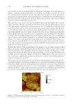
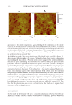
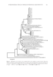
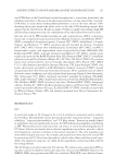
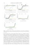
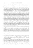
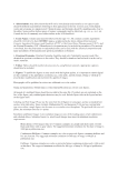
INVESTIGATION OF THE INTERNAL STRUCTURE OF HUMAN HAIR 123 structure observed for the cuticle was not found in the cortex region. Furthermore, the granular nature of the cuticle was proposed by Powell and Rogers to arise because of cysteine-rich granules from the cytoplasm that are fused together during the formation of the mature hair shaft (44). The hair cortex is comprised of elongated cortical cells approximately 5 μm in diameter and 50 μm in length. The hierarchical appearance of the intermediate fi laments can be described by envisioning two strands of slightly different keratin proteins arranged in an in-phase fashion and twisted together to produce a two-stranded rope (a coiled coil). Pairs of these associate to produce an antiparallel offset protein tetramer. By a process, the fi ne details of which remain to be elucidated, the tetramers associate both longitudinally and laterally to produce a discrete rod of semi-infi nite length containing 32 protein chains in its transverse section. This is the “crystalline” part of the hair fi ber known as the keratin intermediate fi lament (or microfi bril). Hundreds of intermediate fi laments are then em- bedded in a pseudo-hexagonal array within a cysteine-rich protein matrix (keratin associ- ated proteins) to form the keratin macrofi bril. Several tens of macrofi brils are packed longitudinally within each cortical cell. The macrofi brils are separated in places by other cellular components such as the effete cell nucleus, melanin pigment granules and, in places, a thin matrix of protein of low cysteine content (sometimes referred to as “non- keratin”). In a recent study, Kadir et al.(45) provided evidence that the amorphous matrix of hair contains granular structures ranging in size from 2 to 4 nm. On cl ose inspection of the cortex region in the AFM micrograph furnished in Figure 3, it is apparent that a number of intricate structures are present. The dark crater-like features (indicated by the white arrows), approximately 0.2–0.5 μm in diameter, more than likely are ghost melanin granules. Possibly, melanin granule structures were present in these locations, but during the hair cross section sample preparation procedure, these were sheared from the exposed surface. Previous studies report melanin granule dimensions on the order of 0.3–0.6 μm in width and 0.8–1.2 μm in length, which are in agreement with the ghost melanin granule measurements (46). We are also able to discern cortical cell structures in Figure 3. These have varying forms and dimensions and are highlighted by the white circles in the micrograph. At times, Figur e 3. AFM micrograph of the cortical region of a cross section of virgin hair. The white arrows indicate places where melanin granules were present, whereas the white circles correspond to cortical cells. The smaller faint blue circles highlight the boundaries of several macrofi brils. It is suspected that nuclear remnants are also present (white asterisks).
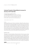
Purchased for the exclusive use of nofirst nolast (unknown)
From: SCC Media Library & Resource Center (library.scconline.org)