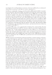
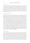
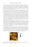
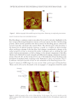
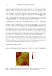
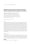
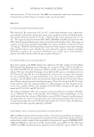
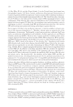
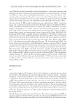
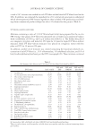
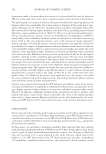
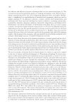
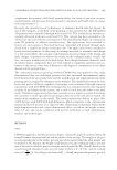
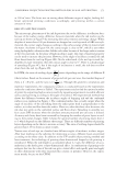
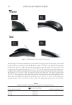
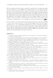
JOURNAL OF COSMETIC SCIENCE 124 cortical cells can have an irregular shape as illustrated in the fi gure. The small features in Figure 3, outlined in blue, correspond to macrofi brils. At a higher resolution, we can see the clear delineation between several macrofi brils (see Figure 4). Each macrofi bril, measured to be approximately 400 nm in diameter, is surrounded by a thin boundary. It has been suggested in the past that there is a demarcation between adjacent macrofi brils denoted as intermacrofi brillar material (47). Most reference texts report that the macrofi bril dimensions for human hair typically range from 40 to 500 nm in diameter (43,48,49). In wool, there are three types of cortical cells: paracortical, orthocortical, and mesocortical cells. In human hair, researchers have used terminology such as ortho-type and para-type cortical cells to denote the similarity of cell types that share some common features with those from wool. The size and struc- ture of macrofi brils in wool are fairly consistent for each cell type. However, the macrofi - brils in human hair were reported to vary considerably in size and shape (50). Therefore, it should come of no surprise that there is such high variability in the size of macrofi brils reported from various sources. Regardless, our measured values (approximately 400 nm) appear to be in line with those previously reported in the literature. Noteworthy, in a study carried out by Harland et al., (47) the thickness of the cross section was found to cause some variability in the measurement of macrofi brillar dimensions because of the tilt angle of intermediate fi laments. Another key feature of hair morphology is the medulla. It is not always present in hair fi bers and is most commonly found in hair of Asian descent. For example, in Caucasian hair, the medulla is not continuous along the length of the fi ber, so depending on the location from which the fi ber cross section is obtained, you may or may not observe the medulla structure. Regardless, Figure 5 contains an AFM image of the cross section of hair containing a medulla. In agreement with previous studies, the medulla appears as a porous and rough structure (51). Historically, not that much attention was given to understanding the structure and func- tion of the medulla. It was merely thought to be a vacuolated morphological component of the fi ber. Studies in recent years, however, have shown that it is rich in lipids and also contain fi brillar protein structures (35,51,52). Furthermore, studies have demonstrated that the medulla is a fairly complex structure and consists of a medullary tube surrounded by two types of medullary cells and vesicles with proteinaceous granules (53). Figure 4. AFM micrograph of a magnifi ed section of the region shown in Figure 3. The slightly elliptically shaped feature in the center of the image is a macrofi bril.

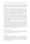
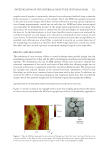
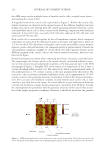
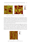
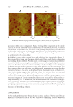
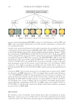
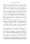
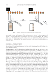
INVESTIGATION OF THE INTERNAL STRUCTURE OF HUMAN HAIR 125 EFFECT OF BLEACHING ON HAIR MORPHOLOGY Hair bleaching is a common cosmetic procedure that is used to introduce highlights to the hair. A typical bleaching formulation contains a two-part system the fi rst is the bleaching powder, which contains persulfate salts that accelerate the bleaching process, whereas the second is typically a developer and contains H2O2. The ultimate goal of this procedure is the destruction of melanin granules however, it results in a number of other secondary effects resulting in damage to the hair fi ber. Such damage is manifested in changes to the surface properties of hair, as monitored by surface techniques such as combing force and surface tension measurements, as well as damage to the internal components of the fi ber, which can be followed by tensile strength and differential scanning calorimetry measure- ments of hair (54,55). At the chemical level, one of the hallmarks of hair bleaching is the breakdown of disulfi de bonds, resulting in the formation of cysteic acid residues (56,57). In addition, the lipid structures in hair are also vulnerable to the bleaching process (58). Figure 6 contains an AFM image of a cross section of the cuticle region of bleached hair. Three cuticle cells can be discerned in the micrograph. A comparison should be made Figure 5 . AFM micrograph of the medulla region of virgin hair illustrating its rough and porous nature. Figure 6. AFM micrograph of the cuticle of bleached hair. In the image of the cuticle, the white arrows indicate regions of the cuticle where severe damage has been induced in the cell membrane complex and endocuticle.
Purchased for the exclusive use of nofirst nolast (unknown)
From: SCC Media Library & Resource Center (library.scconline.org)