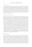
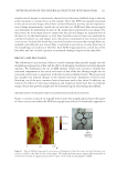
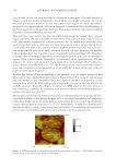
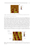
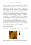
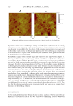
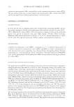
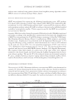
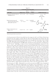
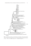
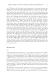
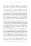
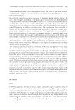
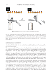
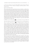
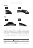
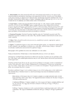
JOURNAL OF COSMETIC SCIENCE 126 with the corresponding image of the cuticle of virgin hair in Figure 2. Structurally, the most noticeable difference between the virgin and bleached hair is the appearance of dark voids that are especially evident in the endocuticle region. Because the endocuticle has the lowest density of crosslinking of the sublamina layers, it seems likely that this would be the region most susceptible to damage from the bleaching procedure. There are also voids in the cuticle of the virgin fi ber (Figure 2) however, they are not nearly as large or deep as those found in bleached hair. These fi ndings are in agreement with TEM studies where it was shown that the electron density of the endocuticle of bleached hair—fi xed with osmium tetroxide and stained with uranium acetate and lead acetate—was greater than that of virgin hair, more so than the other lamellar structures (59). An AFM micrograph of the cortex of bleached hair is provided in Figure 7. Immediately apparent in the image are the large amounts of cracks, crevices, and other asperities in the bleached hair cross section (compare with virgin hair shown in Figure 3). Focusing in on the cortex at greater resolution allows us to discern the macrofi brillar structures and to make a comparison between virgin and bleached hair (Figure 8). In the bleached sample, there appears to be an erosion of biomaterial at the outer edges of the macrofi brils. This could suggest the presence of a low-sulfur–containing protein more susceptible to degra- dation by bleaching than the inner core of the macrofi bril, or the possibility that a cell membrane-like material could be present on the exterior of the macrofi bril and then removed by the bleaching procedure. It should also be pointed out that, regardless of the material, erosion would be expected to proceed from the exposed edges inward. We did not observe noticeable differences in the medulla region of bleached hair as com- pared with virgin hair. This is surprising because previous studies demonstrated greater quantities of lipids in the medulla than other regions of the fi ber (52). Removal of lipids by bleaching and other chemical processes could result in a change in the morphological appearance of the medulla, although this requires further investigation. INVESTIGATION OF DELIPIDATED HAIR Hair contains a variety of lipids that consist of free fatty acids, cholesterol, ceramides, cholesterol esters, and cholesterol sulphate (60). Typically, hair lipids are categorized as Figure 7. A FM micrograph of the cortex of bleached hair. The arrows in the image of the cortex point to major cracks and crevices within the cortical structure caused by the bleaching procedure.

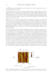
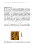
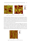
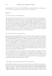
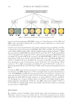
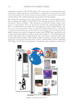
INVESTIGATION OF THE INTERNAL STRUCTURE OF HUMAN HAIR 127 endogenous or exogenous. Endogenous lipids carry out a structural function, serving as the building blocks of the cell membrane complex in hair. Compositionally and structur- ally, there are three different types of cell membrane complex in hair fi bers: (i) cuticle cell membrane complex situated above and below each cuticle cell, (ii) cortex cell membrane complex that surrounds each spindle-shaped cortical cell, and (iii) cuticle–cortex cell membrane complex that provides the boundary between the cuticle and cortex (61). Exogenous lipids arise from the sebaceous gland, which coats the exterior of the fi ber and also secretes lipids within the fi ber structure. The Soxhlet extraction method used in this study more than likely removes both endog- enous and exogenous lipids leaving only covalently bound 18-MEA, which is present in the outermost layer of each cuticle cell. Figure 9 contains an AFM micrograph of the cuticle of delipidated hair. It appears there is a greater degree of demarcation between cuticle cells than the virgin hair (see Figure 2). In addition, the cells adopt a swollen appearance—similar to that found in an earlier TEM study by Takizawa et al. (62) in permanent waved hair. The density of the various lamellar structures is less distinct in the delipidated hair than virgin and bleached hair. Overall, there appears to be a less granular Figure 8. A F M micrographs of the cortex of (A) virgin hair and (B) bleached hair. Figure 9. AF M micrograph of the cortical region of delipidated hair.
Purchased for the exclusive use of nofirst nolast (unknown)
From: SCC Media Library & Resource Center (library.scconline.org)