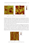
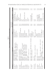
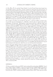
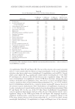
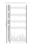
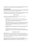
JOURNAL OF COSMETIC SCIENCE 138 Table III List of Primers Used in the MLST Locus Amplifi cation Gene name Size (bp) of fragment analyzed Locus primer (5′→3′) Locus primer (5′→3′) atpD 443 GTTCATCTGGCCGTACAC AACTGACGCTCGAAGTCC gltB 400 CTTCTTCTTCGTCGCCGA TTGCCGACGTAGTCGTTG gyrB 454 ATCGTGATGACCGAGCTG CGTTGTAGCTGTCGTTCC recA 393 TGACCGCCGAGAAGAGCAA GACCGAGTCGATGACGAT lepA 397 GGCATCAAGGAACTGACG CTGCGGCATGTACAGGTT phaC 385 AGACGGCTTCAAGGTGGT ACACGGTGTTGACCGTCA trpB 301 CTGGGTCACGAACATGGA CCGAATGCGTCTCGATGA analyses were conducted using genetic-distance–based neighbor-joining algorithms within MEGA version 4.0 software package (Tokyo, Japan). MLST LOCI AMPLIFICAT ION AND SEQUENCING MLST was performed b y sequencing the following housekeeping genes: ATP synthase beta chain (atpD), glutamate synthase large subunit (gltB), DNA gyrase subunit B (gyrB), recombinase A (recA), GTP-binding protein (lepA), acetoacetyl-CoA reductase (phaC), and tryptophan synthase subunit B (trpB) according to the previously published method available at www.pubmlst.org/Bcc. Primer sequences used for the MLST locus amplifi ca- tion are listed in Table III. Genomic DNA was isol ated using the genomic DNA extraction kit (TaKaRa) mentioned previously, according to the manufacturer’s instructions for bacterial cells. Amplifi cation of MLST loci was performed in 50 μl PCR volumes containing 2× EasyTaq® PCR Super- Mix (TransGen Biotech, Beijing, China), 0.4 μM primers, DNA templates, and double distilled water. Amplifi cation was performed with the Eppendorf Mastercycler® 5,330 using the following cycling conditions: initial denaturation for 2 min at 94°C followed by 30 cycles of 1 min at 94°C, 30 s at an annealing temperature of 58°C, and 2 min at 72°C, followed by a fi nal extension step of 5 min at 72°C. The reaction products were separated and detected on an ABI PRISM Genetic Analyzer 3100 (Applied Biosystems). The sequences from both strands of a given locus of the same isolate were aligned, trimmed to the desired length, and edited using the SeqMan II program from the Laser- gene software package. The specifi c ST of the analyzed strains was determined using the B. cepacia complex multilocus sequence typing website (http://pubmlst.org/Bcc/) devel- oped by Keith Jolley at the University of Oxford (England, United Kingdom) (39). ANTIMICROBIAL SUSCEPTIBILITY T ESTING Determination of the MIC. Mini mum inhibitory concentrati ons (MICs) were determined on a panel of 25 strains illustrating the diverse origins of the collection, broth microdilution using 96-well microplate. First, preservative mother solution was prepared according to the concentrations from Table IV and diluted 10 times, and then a double dilution up to six different dilutions (Table IV) was made. For each bacterial isolate to be tested, 100 μl

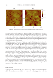
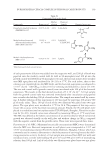
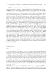
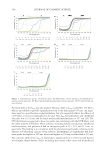
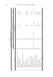
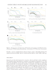
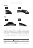
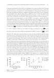
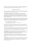
BURKHOLDERIA CEPACIA COMPLEX IN PERSONAL CARE PRODUCTS 139 Table IV Compounds Used in this Study Compounds Range tested Mother solution Chemical formula Chemical structures Dimethoxy dimethyl hydantoin (DMDMH 0.6%)a 0.0125–0.4% 4.0% C7H12N2O4 Methylisothiazolinone and chloromethylisothiazolinone (MIT/cMIT 0.0015%)a 1.5625 × 10⁻⁴–0.05% 0.5% C4H5NOS C4H4NClOS Methyl 4-hydroxybenzoate (MH 0.4%)a 0.0125–0.4% 4.0% C8H8O3 a Maximum permitted concentrations. of each preservative dilution was added into the respective well, and 200 μl of broth was pipetted into the sterility control well (G well in 96-microplate) and 100 μl into the growth control well (H well in 96-microplate). Second, the bacterial isolates were streaked onto MH agar plates and incubated for 18–24 h at 37°C. For each isolate, three to fi ve colonies were transferred into the MH broth, and the bacterial suspension was adjusted to 1 × 10⁸ cfu ml-1 (the OD600 is about 0.1) by vortexing and diluted by a factor of 1:100. The test and control wells (growth control) were inoculated with 100 μl of the bacterial suspension. This results in the fi nal desired inoculum of 5 × 10⁵ cfu ml-1. A 10-μl sample from the growth control tube was removed immediately after inoculation and pipetted into a sterile tube holding 990 μl of sterile saline. The sample was mixed well by vortex- ing. A further dilution of this suspension (1:10) was made by pipetting 100 μl into 900 μl of sterile saline. Then, 100 μl of each of the two dilutions was plated onto two agar plates. The agar plates were incubated at 37°C for 24 h. The purpose of this step was to ensure the accuracy of the bacterial inoculation amount. Finally, the 96-well plates were placed in a multifunctional microplate reader (TECAN Spark, Shanghai, China) for incuba- tion for 24–30 h. Growth curves were obtained by measuring OD600 at 37°C every hour. The MIC was defi ned as the lowest concentration of an antimicrobial at which no bacterial growth was observed visually on the well plate and without change in OD600 compared with the negative control from the growth curves. These tests were repeated three times. Determination of the MBCs. Minimum bactericidal conce ntrations (MBCs) were dete rmined after the MIC test. Bacterial suspensions in well plates were resuspended at 1/10e and 1/100e in neutralizing solution (Fischer Scientifi c Bioblock, Shanghai, China), which was commonly used in cosmetics laboratories to check the presence of surviving bacteria by inhib- iting preservative activity. In detail, a 10-μl sample was removed from MIC test wells and introduced into a sterile tube holding 90 μl (1/10e) and 990 μl (1/100e) of neutralizing solution, then 100 μl and 1 ml of each of the two dilutions were plated onto two agar plates
Purchased for the exclusive use of nofirst nolast (unknown)
From: SCC Media Library & Resource Center (library.scconline.org)