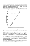
J. Soc. Cosmet. Chem., 33, 1-7 (.January/February 1982) Quantitative aspect of corneocytes P. CORCUFF, G. DELESALLE and H. SCHAEFER, Laboratoire de Recherche, L'Oreal, I ave. de Saint-Germain, 93601 Aulnay-sous- Bois, France. Received March 28, I98I. Synopsis A novel technique for harvesting corneocytes has been evaluated on forearms of 20 volunteers. This procedure removes horny cells with minimal frictional effect on the skin surface and is proposed to evaluate the ease of desquamation. Harvested corneocytes are stained and counted manually or automatically, in "Nageotte" cells. The automatic counting was carried out by an Image Analyser (Quantimet 720) and good correlation with the manual measurements (R 0.99) was obtained. In addition to rapid counting, the Quantimet allows for the evaluation of the surface area of cells and for estimation of the number of broken cells or aggregates. About 10,0OO corneocytes per cm 2 are removed using this procedure. Compared to other techniques, this number is higher than with the passive method but lower than with the scrub technique used by R. Marks et al (2). INTRODUCTION In 1969, McGinley (1) described a scrub technique using a non ionic detergent and obtained measurements of individual cells from the outermost layers of the human stratum corneum. Eight years later, S. Nicholls and R. Marks (2) automated the technique with a "scrub apparatus" which permits the count and some cytomorphological observations (3-6) of the corneocytes. However, this method is a two step process which is time consuming (10, 11). Also, because of the frictional effects, the scrub technique is not a measure of natural desquamation, but of a "forced desquamation" related to cohesive forces between squames (6). Other techniques such as Methocel Gel (7) or ultrasonics (8), are inadequate for morphological measurements because they modify the aspect of harvested features (aggregates, broken cells, shape of corneocytes, extra features). The procedure described in this paper is fully automated and allows for evaluation of natural desquamation, counting and morphological measurements on a large number of cells in a short time.

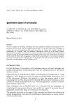
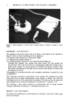
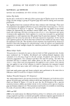
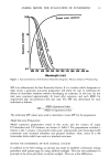
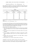
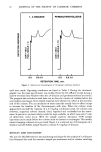
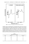
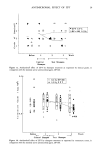
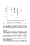
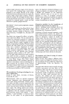
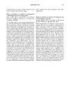
2 JOURNAL OF THE SOCIETY OF COSMETIC CHEMISTS . 5 Figure 1. Turbine apparatus. 1--electric motor 2--plastic syringe 3--chamber 4--support, 5--power supply. MATERIALS AND METHODS The apparatus used in this study is shown in Figure 1 with details of the chamber in Figure 2. There are three essential parts of our apparatus: 1) an electric motor driven by a 5 volts power supply 2) a stainless steel propeller (Figure 2) 3) a cylindrical plastic chamber of 2-cm internal diameter (Figure 2). Through this chamber, two cavities have been bored: The first permits the introduction of three ml of 0.1% triton X-100/phosphate buffer pH 7.9 mixture by a plastic sterile syringe. The second prevents air bubbles entering the chamber. Once the turbine is positioned on the body site, the extent of skin surface in contact with the solution is 3.14 cm 2. The propeller is 5 mm above the skin, and in standard conditions, is rotated for ! min at 2,500 R.P.M. The corneocyte suspension is reaspirated via the syringe, then transfered into glass tubes and 50 /xl of basic fuschin/crystal violet mixture is added. The tubes are centrifuged and 2 ml of the supernatant is discarded. The tubes are shaken vigorously and an aliquot of the corneocyte suspension is introduced to "Nageotte" cell. The counting is done either manually or automatically with an Image Analysis Computer: Quantimet 720 (Imanco--Cambridge Instrument Co.). RESULTS AND DISCUSSION All studies were performed on volar forearms of volunteers, males and females, aged from 25 to 60 years.
Purchased for the exclusive use of nofirst nolast (unknown)
From: SCC Media Library & Resource Center (library.scconline.org)