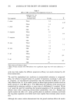
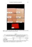
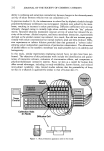
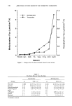
SKIN PENETRATION 233 EXPERIMENTAL MATERIALS The solutes utilized were methylparaben, propylparaben (Fisher Scientific Company, Fairlawn, NJ) and theophylline (Eastman Kodak Company, Rochester, NY). Solute melting points and HPLC chromatograms were used to establish purity. The solvents were distilled water, spectrograde alcohols, propylene glycol, polyethylene glycol 400 (PEG 400), glycerin (Fisher Scientific), and dimethylisosorbide (ICI Americas, Wil- mington, DE). EQUIPMENT A teflon flow-through cell mounted into a temperature controlled aluminum block (Crown Glass, Somerville, NJ) was used in the permeation studies (Figure 1). The body portion of the cell contained the inlet and exit ports and a glass window allowing inspection for air bubbles. Prepared skin was mounted stratum corneum side up in the body of the cell. A cylinder was locked into the cell body (using a threaded ring) and formed the donor compartment. The cylinder was modified by increasing its length in order to reduce skin torque during mounting and to increase donor compartment volume. The area available for diffusion was 0.64 cm 2. Receptor solution was delivered to the cells via a peristaltic pump, and effluent was collected into tared vials. A fraction collector allowed the unattended collection of samples at specified time intervals. METHODOLOGY Male fuzzy rats (Skh:fz Temple University, Philadelphia, PA), 10-weeks-old, weighing 275-300 g, were used in this study. The animals were sacrificed by CO2 asphyxiation. Excised skin samples were stored in a freezer for less than eight weeks. The samples were sufficiently thawed before use, and the dorsal region was removed and dermatomed to a thickness of 320 !xm or 450 !xm (Pagett Dermatome, Kansas City, MO). After gross examination to eliminate damaged skin specimens, the skin was then mounted into the diffusion cell as previously described. The cells were heated to main- tain a 37øC temperature at the dermal side of the skin. The receptor solution was the same throughout the study and consisted of normal saline with 0.25% w/v chlorobu- tanol. The receptor reservoir was maintained at 40øC to reduce formation of air bubbles under the dermal surface. Entrapped air was removed by careful tilting of the cell. Receptor fluid was pumped through the cell at a controlled rate to allow detection of Table I Inter- Versus Intra-Subject Variation in Flux for Aqueous Suspensions of Theophylline (450-•m Skin Thickness) Flux - SD (Ixmol/cm2/h) cv (%) Within an animal (n = 6) Between animals (n = 6) 0.12 -4- 0.02 0.14 + 0.01 12 10

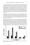
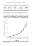
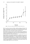
234 JOURNAL OF THE SOCIETY OF COSMETIC CHEMISTS Table II Influence of Skin Thickness for Saturated Aqueous Systems Theophylline Methylparaben Thickness Flux _+ SD Lag time -+ SD Flux + SD Lag time -+ SD (Ixm) (mg/cm2/h) (h) (mg/cm2/h) (h) 320 0.024 + 0.003 1.8 + 0.7 0.11 _+ 0.02 2.9 + 0.7 450 0.024 + 0.003 1.2 +- 0.5 0.096 + 0.02 3.0 -+ 0.3 permeant (approximately 3 ml/h). The mounted skin was allowed to equilibrate for one hour before application of the donor solution. The donor compartment was sealed with parafilm to eliminate evaporative loss. Effluent samples were collected periodically and weighed to determine the actual amount of solution delivered. Samples were filtered through a 0.45 !xm filter, and 20 !xl were injected onto a reversed phase HPLC column (Zorbax ODS-3 column, Dupont, Wilmington, DE) with UV detection-theophylline at 272 nm, parabens at 254 nm. 0 0 20 40 60 80 100 120 Time (h) Figure 2. The cumulative amount of theophylline penetrating the excised skin sample versu• time from infinite aqueous dosing.
Purchased for the exclusive use of nofirst nolast (unknown)
From: SCC Media Library & Resource Center (library.scconline.org)