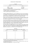
250 JOURNAL OF THE SOCIETY OF COSMETIC CHEMISTS they suggested was hydrogen-bonded to the protein-bound water. Above 50% water content, the water resembled the bulk liquid. The efficacy of skin moisturizers in vivo is usually determined by indirect measurements of hydration. Tests include high frequency electrical conductivity, TEWL (transepider- mal water loss), biomechanical measurements, and subjective clinical evaluations. All suffer from low precision and no well-understood relationship to water content. Spectroscopic measurements, on the other hand, are related to water content in a straightforward manner through a classical Beer's Law type relationship via absorption of the hydroxyl moieties. The absorbance of a sample is proportional to both concen- tration and the path length that light travels. In the mid-infrared, ATR (attenuated total reflectance) has been used to measure water content of skin in vivo. The ratio of the amide I to the amide II band of skin protein has been used as a measure of water content by exploiting the overlap of the water-bending mode with the amide I band (5). This method assumes that neither the amide I nor amide II band is affected by water content, an unproven assumption. Potts et al. (6) have used the 2100 cm- 1 combination band of water, which has the advantage of being far removed from bands due to skin or products. ATR measurements, however, require occlusion of the skin, which can affect water content. In addition, the degree of contact between the skin and the internal reflectance element is not constant, particularly after skin is treated with a lotion, and the refractive index of skin may change upon hydration, thus affecting the depth of penetration of radiation. In this paper, near-infrared reflectance (NIR) spectroscopy is used to determine skin water content in vivo. NIR has several advantages over mid-infrared spectroscopy. The near-infrared region is more sensitive to hydrogen-bonding differences and thus can distinguish different types of water scattering effects can be used to determine changes in the character of the skin surface, and non-occlusive measurements can be made. The possibility of using NIR for this purpose was previously demonstrated in our laboratory by Walling and Dabney (7). In those experiments, a 10-foot fiber optic cable was coupled to the spectrometer to collect spectra on the skin of legs treated with moisturizer. In situ experiments on pigskin distinguished free and bound water, al- though in vivo experiments did not. This paper presents in vivo results determined on skin without a fiber optic cable. The improved resolution allows several types of water, including free, bulk, and protein-bound, to be distinguished. EXPERIMENTAL Dry leg studies were conducted on 16-22 female subjects in successive clinical trials. Four to five sites on the outer lower legs, each about 2 inches square, were used. An untreated control site was included and used to study the effects of ambient humidity on skin water content. The effects of three formulations used in a single trial were compared by NIR. These formulations were 1) a plasticizer (the exact nature of which is proprietary) in an aqueous gel base containing hydroxyethyl cellulose and preserva- tives, 2) propylene glycol (a penetration enhancer) in the same gel base, and 3) a non-ionic oil-in-water emulsion containing 22% oil phase and a water phase that includes the plasticizer, dipropylene glycol, glycerol, and emulsifiers. Product appli- cations were made twice a day for two to four weeks, followed by a regression period of

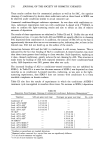
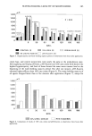
MOISTURE MEASUREMENT BY NIR SPECTROSCOPY 251 no treatment. NIR reflectance measurements were made prior to treatment and at least once a week for the duration of the study. Ambient temperature and relative humidity were not closely controlled but were monitored over the measurement period. Reflectance measurements were made with a Quantum 1200 analyzer (LT Industries) in the region of 1200-2400 nm, using 30 scans per spectrum. The subjects' legs were placed directly against the detector port, with a foam cushion around the rim of the port to block stray light. The spectrometer was placed on a low stool, and subjects could easily reach the detector port from a sitting position. Spectra were collected at least eight hours following the most recent application to ensure that no residual product was present. DATA TREATMENT The spectra were converted to apparent absorbance (log l/R, where R is reflectance) and averaged over all the subjects for each product at each time point. Averaging the data in this manner minimizes the effect of individual variation by limiting the influence of outliers. The averaged spectra within a study were subjected to a multiplicative scatter correction (MSC) to correct for the changes in apparent absorption due to scattering. MSC was first proposed by Geladi et al. for determinations of meat composition (8) and has since been used in a variety of NIR applications (9). MSC corrects each spectrum to an "ideal" spectrum, in this case an average spectrum, and determines both additive and multiplicative scattering effects. MSC assumes a constant scattering coefficient over the wavelength interval used and only minor changes in concentration. Over the wavelength interval used here, 1400-2200 nm, the scattering coefficient of skin has been shown to be fairly constant (10,11), and the variation in spectral intensity is small. From the MSC-corrected data, concentration changes can be determined, and from the difference in the original and MSC-corrected spectra, changes in scattering can be determined. Relative concentrations were determined from second derivative intensities. Differenti- ation is a common means of resolution enhancement, and because it is a linear operation, the resulting spectral intensities follow Beer's Law if it held for the original spectrum (12). RESULTS BAND ASSIGNMENTS The log 1/R spectra of skin and water are compared in Figure 1. The major features in both are the bands near 1450 and 1920 nm, corresponding to the first overtone of the OH stretch in water and the combination mode of the OH stretch and HOH bend in water, respectively. Skin also shows weak features near 1730 and 1750 nm due to lipids. The features in the water combination region are enhanced using second derivatives, as shown for both water and skin in Figure 2. Water shows a major band at 1892 nm and weaker bands at 1906 and 1924 nm. Longer wavelengths generally indicate greater hydrogen bonding. According to Luck, these wavelengths correspond to unbonded (free) water and water with one and two hydrogen bonds, respectively (13). The same three bands appear in skin, with different relative
Purchased for the exclusive use of nofirst nolast (unknown)
From: SCC Media Library & Resource Center (library.scconline.org)