266 JOURNAL OF THE SOCIETY OF COSMETIC CHEMISTS The above radiolabeled compounds were diluted in their non-radioactive counterparts so that the final activities were between 0.1 and 0.3 microcuries per gram of solution. Wool swatches (7.6 x 11.4 cm wool challis, Test Fabrics, Inc., Middlesex, NJ) were prepared for radiotracer experiments by overnight immersion in a 1:1 mixture of acetone and ethanol. Following overnight drying, the cleaned swatches were cut with pinking shears into 0.14-g segments having areas of approximately 8.4 cm 2. At least five of the cut segments were prepared for each treatment under study. In all experiments, cut swatches were wetted with tap water, excess water squeezed out, and the swatch laid on a stainless steel wire gauze placed over a beaker. Following this, either 0.3 or 0.15 ml of a test material was applied to the swatch as evenly as possible. The wool was then rubbed for one minute between two lengths of rubber tubing mounted on stainless steel rollers especially constructed in our laboratories. After rubbing, swatches were rinsed in a beaker of tap water for 45 seconds, followed by a 15-second rinse in a second beaker. After the latter rinse, very little radioactive material could be removed from the swatches and a final rinse was performed for one minute under 100 ø F running tap water. Depending upon the particular experiment, after the above treatment, wool swatches were either hung up to dry overnight or treated again after excess water was squeezed out using rubber tubing placed over the ends of crucible tongs. Following treatment and drying, wool swatches were dissolved by placing each in a glass counting vial, adding 1 ml of 2M NaOH, and heating in an oven at 80 ø C for 1.5 hours. Three reference vials were also prepared in which a wool swatch was dissolved following addition of a known amount of radioactive material. After cooling, approximately 12 ml of Aquasol-2 LSC cocktail was added to each vial, followed by addition of 0.25 ml of concentrated perchloric acid. The vials were then shaken vigorously, resulting in a clear solution ready for counting. All measurements with radiotracers were run using only one radiolabeled substance at a time. Thus, for example, in experiments in which both conditioner and detergent deposition were measured for a given treatment series, the series was run twice, once with radiolabeled detergent and once with radiolabeled conditioner. LIGHT-SCATTERING MEASUREMENTS The use of a goniophotometer to carry out light-scattering measurements was described in reference 1. In the current work, all goniophotometric measurements were performed using a Murakami GP-1R automatic goniophotometer purchased from Hunter Associ- ates Laboratory, Reston, VA. The GP-1R, which employs collimated light from a tungsten halogen lamp, has several advantages over the Brice-Phoenix photometer employed in the previous work (1). Among these are the fact that it is a double goniometer instrument so that the sample can be turned to any angle desired with respect to the incident light. In addition, it has a large sample compartment permitting the sensitive stepping motors to run scans from 0 ø to 75 ø (or a full 360 ø if so desired). Finally, the instrument accuracy is quite high, with a specified scan repeatability of ---0.2%, an angle accuracy of ---20', and a pho- tometric accuracy of ---0.5 %.

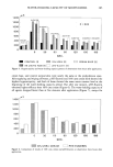
SURFACTANT INTERACTIONS 267 As in the previous work (1), for all light-scattering experiments, polarizers having directions of polarization perpendicular to the plane of incidence were placed between the light source and the sample and between the sample and the light detector of the goniophotometer. In the current experiments, a Glan-Thompson prism was used in front of the sample, while a sheet polarizer was placed before the light detector. Figure la shows the bottom plate of the GP-1R sample holder used for mounting hair fibers. The diameter of the opening in this plate is 7.3 cm, while the raised plastic ring is 2-mm high and 2-mm thick. In order to accommodate up to 40 hair fibers, the plastic ring contains 40 grooves, 0.635 mm apart, cut at opposite ends of the ring. In all experiments in this work, only one hair fiber was used per scan. These fibers were mounted on the bottom plate under the constant tension supplied by a 2-g alligator clip and held in place by a cover plate secured with two strips of magnetic tape. Figure lb shows the complete sample holder with a single fiber mounted. All tress measurements were made by taking 21 hairs from a treated tress, scanning Figure l a. Photograph of the bottom plate of a GP-1R hair sample holder. Note the four circular magnets set in the bottom plate and the two holes on either side. These are used to mount the sample holder on the vertical mount in the light-scattering instrument.
Purchased for the exclusive use of nofirst nolast (unknown)
From: SCC Media Library & Resource Center (library.scconline.org)