182 JOURNAL OF COSMETIC SCIENCE (20) (21) (22) (23) (24) (25) (26) (27) (28) (29) (30) (31) (32) (33) (34) (35) (36) (37) (38) (39) (40) (41) G.J. Smith, R. F. C. Claridge, and C.J. Smith, The action spectra of free radicals produced by the irradiation of keratin containing bound iron(III) ions, Photochem. Photobiol., 29, 777-779 (1979). K. D. Goddinger and H. Hi3cker, Photodegradation of tryptophan in wool,J. Soc. Dyers Colour., 113, 350-355 (1997). R. F. Borkman, Ultraviolet action spectrum for tryptophan destruction in aqueous solution, Photochem. Photobiol., 26, 163-166 (1977). W. Korytowski, B. Pilas, and T. Sarna, Photoinduced generation of hydrogen peroxide and hydroxyl radicals in melanins, Photochem. Photobiol., 45, 185-190 (1987). T. Sarna, A. Duleba, W. Korytowski, and H. M. Swartz, Interaction of melanin with oxygen, Arch. Blochem. Biaphys., 200, 140-148 (1980). X. Qu, L. J. Kirschenbaum, and E. T. Borish, Hydroxyterephthalate as a fluorescent probe for hydroxyl radicals: Application to hair melanin, Photochem. Photobiol. 71, 307-3 ! 3 (2000). X. Fang, G. Mark, and C. V. Sonntag, OH radical formation by ultrasound in aqueous solutions. Part I: The chemistry underlying the terephthalate dosimeter, Ultrason. Sonochem., 3, 57-63 (1996). T. J. Mason, J.P. Lorimer, D. M. Bates, and Y. Zhao, Dosimetry in sonochemistry: The use of aqueous terephthalate ion as a fluorescence monitor, Ultrason. Sonochem., 1, S91-S95 (1994). J. C. Barreto, G. S. Smith, N.H. P. Strobel, P. A. McQuillin, and T. A. Miller, Terephthalic acid: A dosimeter for the detection of hydroxyl radicals in vitro, Life Sci., 56, 89-96 (1995). G. R. Buettner and L. W. Oberley, Consideration in the spin-trapping of superoxide and hydroxyl radical in aqueous systems using DMPO, Blochem, Biophys. Res. Cammun., 83, 69-74 (1978). E. Finkelstein, G. M. Rosen, and E. J. Rauckman, Spin trapping of superoxide and hydroxyl radical: Practical aspects, Arch. Biochem. Biophy., 200, 1-16 (1980). S. Persad, I. A. Menon, and H. F. Haberman, Comparison of the effects of UV-visible irradiation of melanins and melanin-hematoporphyrin complexes from human black and red hair, Photochem. Pho- tobiol., 37, 63-68 (1983). T. Sarna and R. C. Sealy, Photo-induced oxygen consumption in melanin systems: Action spectra and quantum yields for eumelanin and synthetic melanin, Photochem. Photobiol., 39, 69-74 (1984). T. Sarna, E. A. Menon, and R. C. Sealy, Photo-induced oxygen consumption in melanin systems. II. Action spectra and quantum yields for pheomelanins, Photochem, Photobiol., 39, 805-809 (1984). R. M. B. Deibel and M. R. Chedekel, Biosynthetic and structural studies on pheomelanin, J. Am. Chem. Sac., 104, 7306-7309 (1982). L.J. Wolfram and L. Albrecht, Chemical- and photo-bleaching of brown and red hair, J. Sac. Cosmet. Chem., 82, 179-191 (1987). E. Hoting, M. Zimmermann, and H. Hi3cker, Photochemical alterations in human hair. Part II. Analysis of melanins,J. Sac. Cosmet. Chem., 46, 181-190 (1995). B. Halliwell and J. M. C. Gutteridge, Free Radicals in Biology and Medicine, 2nd ed. (Oxford University Press, Oxford, 1989), pp. 18-19. G. Suez, P. J. Thornalley, H. A. O. Hill, R. Hems, and J. V. Bannister, The production of free radicals during the autooxidation ofcysteine and their effect on isolated rat hepatocytes, Biachim. Biophys. Acta, 719, 24-31 (1982). K. D. Held and J. E. Biaglow, Mechanisms for the oxygen radical-mediated toxicity of various thiol- containing compounds in cultured mammalian cells, Rad. Res., 139, 15-23 (1994). A. Strasheim and K. Buijs, An infrared study of the oxidation of the disulfide bond in wool, Biochim. Biophys. Acta, 47, 538-539 (1961). V. Signori and D. M. Lewis, FTIR investigation of the damage produced on human hair by weathering and bleaching processes: Implementation of different sampling techniques and data processing, Int. J. Cosmet. Sci., 19, 1-13 (1997).

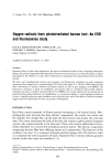
j. Cosmet. Sci., 51, 183-192 (May/June 2000) Near-infrared spectroscopy: Applications in hair research CHANDRA M. PANDE and BRIAN YANG, Bristol-Myers Squibb World Wide Beauty Care, 2 Blachley Road, Stamford, CT 06922. Accepted for publication March 15, 2000. Synopsis We have evaluated the potential of NIR (near-infrared) spectroscopy as a tool in hair research. We find that this technique is ideally suited for measuring the relative moisture content of hair in situ under practical hair-grooming conditions. It also allows measurement of melanin absorption in the NIR region, where there is no interference from synthetic hair dyes. Thus, rapid evaluation of "lift," the bleaching produced by oxidation dye products, can be performed on hair swatches as -well as on live heads. INTRODUCTION Human hair fibers are roughly 50-80 l•m in diameter and are primarily composed of keratin proteins. Nearly 95 % of the dry hair mass is proteinaceous, and 10% of this derives from the disulfide-containing amino acid, cystine. The remaining is made up of lipids, pigments, and some bound ions (1). Under ambient conditions of 25øC and 50% RH (relative humidity), hair fibers bind as much as 10% water by weight (2). Hair fibers have a complex morphology, with three distinct regions: the outermost cuticle layers, the inner cortical cells, and occasionally an innermost and porous medulla. The cortical cells contain or-helical protein, assembled in a fibrillar arrangement, embedded in an amorphous protein matrix (3). Water acts as a plasticizer for hair and plays a critical role in determining its tactile, mechanical, and other cosmetic properties. The amount of water bound to hair depends on the ambient humidity, with more water bound at higher humidities. The affinity for water mainly arises from the polar amino acid side chains of keratin, with negligible contribution from the peptide bonds (4,5). As the humidity increases, one would expect the binding to become less specific, with water binding to low affinity sites as well as existing as free water. The moisture content of hair at any given RH, as measured by the regain from the dry state, is different and less than that obtained by way of dehydrating hair from 100% RH. Such hysteresis is also seen in other synthetic polymers and biopolymers and has been explained to arise from differences in the ratio of the "bound" to the "free" water (6). Watt (2) provides an excellent review of the water-binding properties of keratin, with ample references. Cosmetic hair treatments, either with heating appliances such as a blow dryer and curling iron or with grooming products such as hair fixatives, modify the water-binding ability of hair and, therefore, its physical 183
Purchased for the exclusive use of nofirst nolast (unknown)
From: SCC Media Library & Resource Center (library.scconline.org)