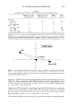
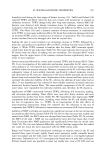
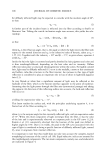
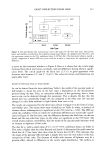
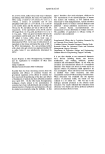
232 JOURNAL OF COSMETIC SCIENCE (sense) to generate biotinylated PCR products detectable by digoxygenin-labeled probes in an immunoenzymatic assay (ELISA) method (17). First, cDNA was mixed with 10x buffer, 10 mM dNTPs, 1 U Taq DNA polymerase (Bioneer, Korea), and iNOS primers in a final volume of 50 lal. The primer sequences and relative predicted PCR product sizes are given below: Human iNOS (sense) 5'-AGTTTCTGGCAGCAACGG-3' Human iNOS (anti-sense) 5'-TTAAGTTCTGTGCCGGCAG-3' A sample containing all reaction reagents except cDNA was used as PCR negative control in any amplification. The mixtures were incubated for the indicated cycles (predenaturation 5 min at 95øC denaturation 50 sec at 95øC annealing 20 sec at 56øC extension 20 sec at 72øC) in a GeneAmp PCR System 2400 (Perkin-Elmer). The correct size of all PCR products was confirmed by comparing with a DNA standard on agarose gel. After a given cycle of PCR, the amount of amplified cDNA was determined by the ELISA method. First, microplates (Maxisorp Nunc) were coated with 50 lag/ml of avidin (Sigma) in coating buffer (CB 15 mM Na2CO3, pH 9.6) and incubated for two hours at 37øC. After incubation, free sites were saturated with 2% blocking solution (Roche, Germany) in CB. Biotinylated PCR products diluted in PBS containing 3% bovine serum albumin (PBSB) were distributed onto microplates (100 lal per well) and incu- bated for one hour at room temperature. After incubation, the microplates were washed three times with PBST. Amplified cDNA was denatured using 0.25 M NaOH at room temperature for ten minutes. Following the washing, 100 lal per well of 10 pmol/ml digoxygenin-labeled probes in hybridization buffer [6.25x SSC, 0.625% blocking re- agent (Roche), 0.125% Tween 20, and 0.5 M NaH2PO 4 (pH 6.5)] were added and incubated at 42øC for two hours. Anti-digoxygenin AP-conjugated antibody (Sigma) was added (1:3000 in PBSB) and incubated for one hour at 37øC. The reaction was developed by nitrophenyl phosphate (pNPP 1M diethanolamine buffer, pH 9.6). The amount of amplified product was measured for optical density at 405 nm (OD 405) using a microplate reader. STATISTICAL ANALYSIS Results were presented as means + standard error (SE). Experimental results were statistically analyzed by using Student's t-test (SigmaPlot 2000). P values 0.05 were regarded as indicating significant differences. RESULTS AND DISCUSSION EFFECT OF UV ON THE PRODUCTION OF MMPS The immunoreactive MMP-1 and -2 in the culture medium of HDFs were measured using anti-MMP-1 and -2 monoclonal antibodies, respectively. Treatment of HDFs with UV radiation enhanced the production of MMP-1 by twofold and MMP-2 by threefold in a dose-related manner (Figure 1), confirming the previous results (1-5). We also confirmed that the gelatinase activities were proportionally increased by the UV irra- diation of HDFs using gelatin zymography (data not shown). The production of MMPs

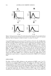
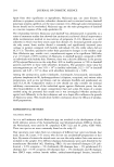
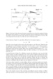
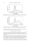
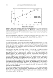
INHIBITION OF MATRIX METALLOPROTEINASES 233 0.6 fl" 0.5 '[] MMP1 •5•. * 0 0.4 :•-• 0 ............ • ........... • .............. • ................ • .... •- ,._t._,_. •! ........ • 0 5 10 15 20 30 UV irradiation (J/c•) Figure 1. The effect of UV irradiation on the production of MMP-1 and MMP-2 by human dermal fibroblasts. HDFs (1.5 x 10⁵/well) were seeded into 35• plates and cultured overnight. The cells were irradiated from a distance of 15 cm by a UV so•ce for a given time. *n = 3. p 0.05 vs no UV ex•sure. by UV irradiation of HDFs is a result of the activation of cell surface growth factors and cytokine receptors, which have in common the requirement for dimerization to initiate signal transduction. UV radiation rapidly activates EGF receptors, followed by the activation of Ras, ERK, JNK, and p38 (18,19). These stress-activated MAP kinases then increase the proteins of c-Jun, c-Fos, and ATF. The dimerization of these proteins activates AP-1 DNA binding and, finally, the induction of MMPs. Fisher et al. showed that retinoic acid can inhibit the induction of MMPs by UV irradiation in human skin by blocking the DNA binding of AP-1 and c-Jun protein induction (20). EFFECT OF NITRIC OXIDE DONOR ON THE PRODUCTION OF MMPS To determine the effect of NO on the production of MMP-1 and -2, HDFs were treated with SNP. SNP is a donor of NO and it can mimic the cellular effects of NO. Treatment with SNP increased MMP-2 production acutely, to about 243% of untreated cells with 50 microM of SNP. The amount of secreted MMP-1 in the HDF culture medium was also increased with SNP treatment, but was less significant than that of MMP-2, about 153% (Figure 2A). The result of zymography also shows that gelatinase activity was increased with SNP treatment. The combined effect of SNP and UV treatments was also tested, as shown in Figure 2B. The null hypothesis was that the effect of UV irradiation is not mediated by NO, and that subsequently the addition of SNP to UV-irradiated HDF should increase MMP production to the same degree as in cells not treated with UV irradiation and more than in cells treated only with UV irradiation. In the UV- treated cells, 50 microM of SNP did not increase MMP-1 production significantly, while that of MMP-2 increased by 196% (Figure 2B). This result indicates that the effect of UV irradiation and SNP treatment on MMP-! production by HDF is not cumulative,
Purchased for the exclusive use of nofirst nolast (unknown)
From: SCC Media Library & Resource Center (library.scconline.org)