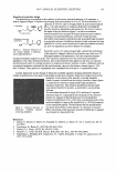
34 JOURNAL OF COSMETIC SCIENCE croplates instead of traditional shake flasks in submerged cultivations. Some targets were isolated by this method (8). The present work reports the isolation and characterization of a novel strain, TI-C3, of Streptomyces hiroshimensis with the highest anti-tyrosinase activity in our screening study. MATERIALS AND METHODS MICROORGANISM Streptomyces hiroshimensis BCRC 12423 used as an indicator for strain identification was obtained from Bioresources Collection and Research Center, Food Industry Research and Development Institute, Taiwan. CHEMICALS Mushroom tyrosinase, L-tyrosine, sodium caseinate, asparagine, sodium propionate, corn steep liquor, maltose, glycerol, sodium nitrate, ammonium sulfate, glucose, nalidixic acid, deoxyribonucleotide triphosphate, and cycloheximide were purchased from Sigma (St Louis, MO). Yeast extract, malt extract, tryptone and agar were obtained from Difeo Laboratories (Detroit, MI). Taq DNA polymerase needed for polymerase chain reaction (PCR) was purchased from Takara Bio (Shiga, Japan). Primers were purchased from MDBio (Taipei, Taiwan). Other reagents and solvents used were commercially available and used as received. IDENTIFICATION OF THE STRAIN TI-C3 The strain TI-C3 was identified according to protocol published by Shirling and Gott lieb (9). The melanoid pigment formation was observed on ISP 1, 6, and 7 media. The carbon source utility was determined on basal medium (Pridham-Gottlieb medium) with 1 % (w/v) of the tested sugar. The basal medium contained 2.64 g (NH4)2SO4, 2.38 g KH 2 PO 4 , 5.65 g K 2 H 2 PO4 • 3H2O, 1.00 g MgSO4 • 7H2O, 6.4 mg CuSO4 • 5H2O, 1.1 mg FeSO 4 • 7H 2 O, 7.9 mg MnCl 2 • H 2 O, 1.5 g ZnSO 4 · 7H 2 O, and 18.0 g agar in one liter of distilled water. The pH was adjusted to 6.9 before autoclaving. The biochemical and physiological characteristics of the strain TI-C3 including growth temperature, melanin production, lysozyme resistance, and substrate hydrolysis were determined by the method of Berd (10). The mycelium of the strain was observed with a light microscope. The spore chain and spore surface morphologies were observed with a scanning electron microscope (Hitachi S-420 Hitachi, Ltd., Tokyo). Cell wall composition (DL- and LL-diaminopimelic acid isomer, A 2 pm) was determined by the method of Hasegawa et al. (11). One or two colonies were placed in a cryogenic vial (Evergreen Scientific) with 0.1 ml of 6 N HCl. The vial was autoclaved at 121 °C for 15 minutes. After cooling, 1 µl of the hydrolysate was placed on a thin cellulose plate (microcrystalline cellulose Tokyo Kasei Co., Ltd, Japan). One microliter of 0.01 M DL-A 2 pm (Sigma) was spotted on the same plate as a standard. The plate was developed on the solvent system methanol-distilled water-6N HO-pyridine (80:26:4: 10, v/v) for three to four hours. The plate was then dried and sprayed with Ninhydrin spray reagent

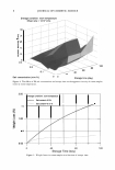
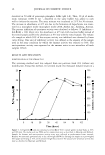
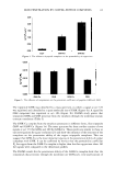
S. HIROSHIMENSIS STRAIN WITH ANTI-TYROSINASE ACTIVITY 35 (Merck) and heated at 100°C for five minutes. The spots of A2pm appeared in a yellowish green color. The sugar content of the cells was analyzed by the same procedure as that of A2pm, but the hydrolysis and developing were carried out by using 2 N trifl.uoroacetic acid and n-butanol-distilled water-pyridine-toluene (10:6:6: 1, v/v) as solvents, respectively. The spraying reagent was acid aniline phthalate. The standard sugar solution contained 1 % (w/v) of each galactose, glucose, mannose, arabinose, xylose, and ribose. The partial gyrB gene fragment was amplified by polymerase chain reaction (PCR) using the primers: PFl (forward: 5 '-GAGGTCGTGCTGACCGTGCTGCACGCGGGCGG CAAGTTCGGC-3') and PR2 (reverse: 5'-GTTGATGTGCTGGCCGTCGACGTCGG CGTCCGCCAT-3'). Genomic DNA was extracted from seven-day cultures using the Qiagen® Genimic DNA Kit. The PCR procedure for amplifying the gyr B sequence was the same as that described by Hatano et al. (12). The amplified product was analyzed in a genetic analyzer (ABI Prism 31 O PE Applied Biosystems, USA) according to the manufacturer's protocol. The gyr B sequence of the strain TI-C3 was aligned manually against the nucleotide sequences of other whorl-forming Streptomyces strains retrieved from GenBank databases. DNA-DNA hybridization was performed by the method of Ezaki et al. (13). The experiment was performed at least five times, and the level of DNA-DNA hybridization was expressed as the mean percent of the homologous DNA binding value. FERMENTATION OF THE STRAIN TI-C3 A seed culture of the strain TI-C3 was initiated by adding 1.5 ml of a thawed spore suspension (about 108 spores per ml) into a 250-ml baffled Erlenmeyer fl.ask containing 25 ml of YMG medium. The compositions of YMG medium are 4 g yeast extract, 10 g malt extract, and 4 g glucose in one liter of distilled water. The pH of the medium was adjusted to pH 7 .2 prior to autoclaving. The fl.ask was then incubated at 30°C for two days at 180 rpm, and 2.5 ml of the seed culture was transferred into a 250-ml baffled Erlenmeyer fl.ask containing 25 ml of modified YMG medium for a secondary cultivation. For fermentation using different carbon sources, glucose in the YMG me dium was replaced by the desired carbon source. For fermentation using different ni trogen sources, both yeast extract and malt extract in the YMG medium were replaced by the desired nitrogen source. All cultivations were carried out at 30°C for 72 hours at 180 rpm. After fermentation, an equal volume of ethanol was added to each cultivation and shaken vigorously for 30 minutes at room temperature. The cell debris was removed by centrifugation at 4800 rpm. The supernatant from the extracted broth was assayed to measure the anti-tyrosinase activity. The experiments were carried out in triplicate and the mean values are shown. ANTI-TYROSINASE ACTIVITY ASSAY The modified assay method of anti-tyrosinase activity described in the literature was employed in our study (14). Each supernatant sample from the fermentation experiments was serially diluted by a reaction buffer before the assays of anti-tyrosinase activity. Then, 20 µl of each of the diluted samples was mixed with 80 µl of 0.2 mM 1-tyrosine
Purchased for the exclusive use of nofirst nolast (unknown)
From: SCC Media Library & Resource Center (library.scconline.org)