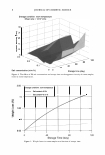
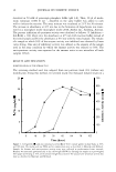
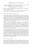
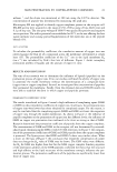
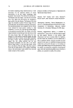
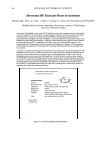
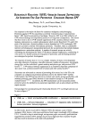
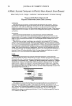
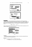
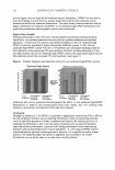
36 JOURNAL OF COSMETIC SCIENCE dissolved in 50 mM of potassium phosphate buffer (pH 6.8). Then, 20 µl of mush room tyrosinase (1000 U ml -1, dissolved in the same buffer) was added to each well to initiate the reaction. The assay mixture was incubated at 25°C for 30 minutes. The increase in absorbance at 475 nm due to the formation of dopachrome was moni tored in a microplate reader (microplate reader 2010, Anthos Inc., Salzburg, Austria). The percent inhibition of tyrosinase activity was calculated as follows: % Inhibition = [(A-B)/A} x 100, where A is the absorbance at 475 nm with reaction buffer instead of the tested sample and Bis the absorbance at 475 nm with the tested sample. The volume of a sample at which 50% of the enzyme activity was inhibited was obtained by linear curve fitting. One unit of inhibitory activity was defined as the amount of the sample used in the assay condition by which the enzyme activity was reduced to 50%. The anti-tyrosinase activity was expressed as the amount units in one microliter of broth samples (U/ml). RESULTS AND DISCUSSION IDENTIFICATION OF THE STRAIN TI-C3 The screening method used was adopted from our previous work (14) without any modification. Using this method, we screened nearly two thousand isolated strains in a 25 20 ■ i 1 5 � 10 u 5 0 0 18 26 30 42 54 Time (hour) 700 600 ◄ 500 400 :E 300 c 200 100 0 78 102 Figure 1. Cell growth(■) and anti-tyrosinase activity (.A) of TI-C3 culture grown in shake flasks at 30°C and 180 rpm. The media used was YMG and its compositions were as described in Materials and Methods. Samples for biomass and anti-tyrosinase activity assay were collected at predetermined time intervals. Biomass was measured gravimetrically as dry cell weight (DCW) by filtering the sample on a pre-weighed filter paper and drying at 70 ° C until constant weight was attained. The assay of anti-tyrosinase activity was done as described in Materials and Methods.

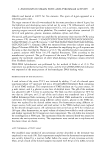
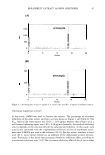
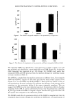
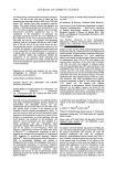
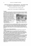
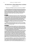
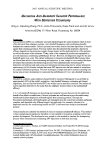
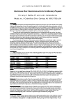
S. HIROSHIMENSIS STRAIN WITH ANTI-TYROSINASE ACTIVITY 37 Figure 2. Light (a) and electron (b) micrographs of the strain TI-C3 grown on an oatmeal agar plate at 30°C for 14 days. Bars represent 10 µm and 0.1 µm in the light and electron micrographs, respectively.
Purchased for the exclusive use of nofirst nolast (unknown)
From: SCC Media Library & Resource Center (library.scconline.org)