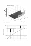
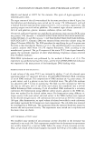
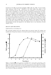
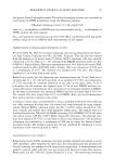
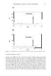
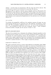
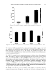
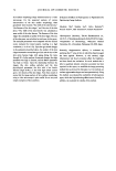
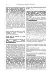
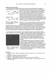
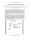
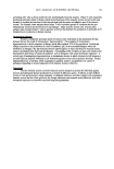
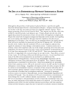
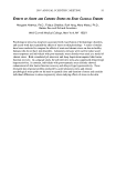
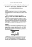
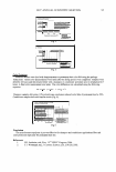
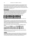
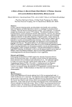
50 "'ii u CJ =a·= tu CD ._ C fl) fl) � � LL. u (I) '?ft. JOURNAL OF COSMETIC SCIENCE 100 -----· .., ... ,' , , I , . , , 50 , , , . I , I , , I � __________.,,,.- _.,,, 0 -+-----...----r------r-------.------, -1 0 1 2 3 Log concentration (µg/ml) 4 - -BHl -- L-ascorbic acid • • • • • A. incisus extract Figure 4. Antioxidant activity of A. incisus ether extract, L-ascorbic acid, and butylated hydroxytoluene. 120 Gi * (J 100 C a, 80 C (J 60 C ·2 40 Gi :l! 20 � 0 0 Control 2 ug/ml 10 ug/mL 15 ug/ml 25 ug/ml Concentration (ug/ml) Figure 5. Inhibitory effects of A. incisus ether extract on melanin synthesis in melanocyte B16Fl melanoma cells. Data are expressed as percent of control, and each column represents mean ± S.D. of triplicate study. Significantly different from the control value: *p 0.5, **p 0.01. skin-lightening agents, kojic acid and hydroquinone. The concentration of each tested sample used was 10 µg/ml, which corresponds to the IC 50 of the ether extract, according to a previous study of tyrosinase-inhibitory activity. Additionally, purified artocarpin, the major component of the A. incisus's heartwood extract, was investigated for its melanogenesis-inhibitory effect. Its concentration used was 4.5 µg/rnl, which is equiva lent to 4.5 µg/ml of artocarpin contained in the extract, according to a previous study of artocarpin content in the extract. The effect of the tested samples on the melanin production of melanocyte B16Fl cells is shown in Figure 6. The reduction of melanin synthesis was expressed by kojic acid, A. incisus ether extract, artocarpin, and hydroquinone as 9.61 %, 8.76%, 8.88%, and 40.97%, respectively, compared to the control cells. These results reveal that, at a similar artocarpin concentration, the inhibition of melanin production of the ether extract shows no difference from that of the artocarpin. Compared to kojic acid, it is likely that the potential of melanin reduction of the ether extract is close to that of kojic acid.

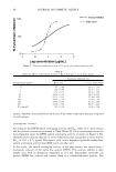
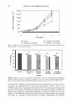
BREADFRUIT EXTRACT AS SKIN LIGHTENER 120 �--------------------------------. 'ii 100 � ! 8 C 'E la 'i :E 80 60 40 � 20 0 Control 10 ug/rrt. Kojic acid 10 ug/rnLA lnclsus Sample * 4.5 ug/rrt. Artocarpln 10 ug/ml. Hydroqulnone 51 Figure 6. Effects of A. incisus ether extract, hydroquinone, kojic acid, and artocarpin on melanin production of melanocyte Bl6Fl melanoma cells. Data are expressed as percent of control, and each column represents mean ± S.D. of triplicate study. Significantly different from the control value: *p 0.5, **p 0.01. Generally, skin pigmentation is determined by melanin synthesis within melanosomes and their distribution to keratinocytes within the epidermal melanin unit. Focusing on melanin synthesis, the rate-limiting step is the hydroxylation of tyrosine to DOPA by the enzyme tyrosinase. Since melanin production requires tyrosine, some mechanism, such as transportation of this amino acid through the melanosomal membrane, may be also a critical factor in limiting pigment production (12,13). As we know, kojic acid, a g-pyrone derivative, can strongly inhibit tyrosinase activity by chelation of the copper ion(s) at the active center of the enzyme (14, 15 ). For artocarpin, as mentioned above, its tyrosinase-inhibitory activity could not be observed. This probably results from its steric hindrance structure since a 4-substituted resorcinol skeleton, coupled with a C3 sub stituent, is present in the flavone structure of artocarpin (16). Therefore, the melano genesis-inhibitory activity of artocarpin may mainly involve the transportation of tyro sine through the melanosomal membrane, a subject that we need to clarify in the future. CELL VIABILITY As an attempt to find out if the reduction of melanin content in B16Fl cell treated with the ether extract causes the reduction of the viable cells, the determination of the effect of such an extract on cell proliferation was performed. B16Fl cells were treated with various concentrations (10, 25, and 100 µg/ml) of A. incisus ether extract for three days, and the number of cells was then determined. The numbers of viable cells were evaluated by staining cells with blue dye. As shown in Figure 7, A. incisus extract induced the dose-dependent inhibition of cell growth in B 16Fl cells. The inhibitory effects on B16Fl cells were less profound at a low dose (25 µg/ml) of the extract, whereas significant arrest of cell growth was observed at a concentration of 100 µg/ml. High doses of the extract may cause cell apoptosis and/or alteration of cell attachment, which is an important characteristic for the proliferation of adherent cells. The effect of kojic acid, A. incisus extract, artocarpin, and hydroquinone on the prolif eration of melanocyte B 16F 1 cells is shown in Figure 8. The obtained results indicate
Purchased for the exclusive use of nofirst nolast (unknown)
From: SCC Media Library & Resource Center (library.scconline.org)