JOURNAL OF COSMETIC SCIENCE 314 they gave their written consent. Two research assistants were responsible for all recruitment and data collection. The in vivo experiments were carried out on the volar forearms of each volunteer, and each subject was rested for 15 min before the experiments and room conditions were set at 22 ± 2 °C and 40–50% relative humidity. Four sites on the ventral surface of each forearm were defi ned using a circular template (1 cm2) and demar- cated with permanent ink. During the fi rst week of experiments, skin tanning was in- duced in each site by exposition of a lamp that simulate sunlight (Helios Italquartz srl, Milan, Italia), which emitted in a range of 300–400 nm (6.5 mW cm−2), for 2–8 min depending on the minimal erythema dose (MED) of each subject. The initial melanin value and the melanin content obtained during the monitoring period were performed by refl ectance visible spectrophotometer X-Rite model 968 (X-Rite Inc., Grandville, MI) as previously reported (21). The variation of skin melanin values was monitored for a total period of 4 weeks. To avoid induced and interfering skin erythema events, exposition to a lamp was not conducted on the third and the sixth day of the fi rst week. For each forearm, three skin sites were treated with tested formulations and the one not treated was consid- ered as control. Tested formulations were applied after each lamp treatment for the fi rst week and once daily in the second, third, and fourth week of the study. To evaluate the time course of skin pigmentation, melanin index (MI) baseline values were subtracted from the MI values to calculate mean MI (ΔMI) values. For each site, plotting ΔMI vs. time, the area under the curve was computed using the trapezoidal rule to obtain area under curve (AUC) dimensionless index values directly related to the degree of skin pig- mentation. From the AUC values obtained, the skin-whitening effect of each formulation was expressed as the inhibition of skin pigmentation (PI) using the following equation: u100 AUC AUC PI (%) AUC C S C (2) where AUCC is the AUC value of no treated skin site and AUCS is the AUC value ob- tained from treated sites. IN VIVO EVALUATION OF PHOTOSENSITIZING EFFECT OF SKIN-WHITENING AGENTS The skin tolerance of skin-whitening agents was investigated by an in vivo model previ- ously reported (21). After a rest period of 6 months, the same subjects participating in the previous study were enrolled to evaluate if skin-whitening application had increased the skin sensitivity to UVB irradiation. For each subject, four skin test sites were defi ned on the ventral surface of each forearm. Three sites were treated with formulations once daily for 4 consecutive weeks and one site was used as control (no topical treatment). At the end of the fourth week, all sites were exposed to UVB irradiation dose, corresponding to the MED by using the lamp previously described to simulate sunlight. The induced ery- thema was measured, after 24 h from the skin site exposure, by refl ectance spectropho- tometry and the registered photosensitivity was expressed as a percentage calculated from erythema index values using equation 3: u100 EI EI Photosensitivity (%) EI T C C (3) where EIC is the erythema index of control sites and EIT is the erythema index of sites treated with formulations.

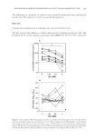
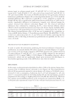
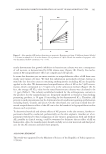
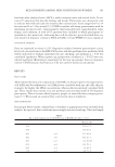
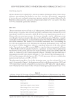
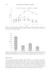
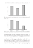
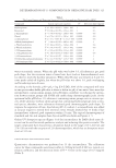
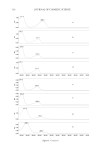
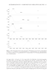
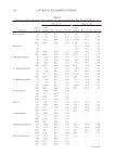
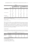
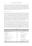
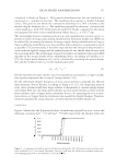
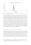
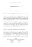
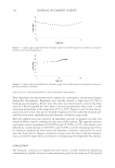
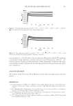
SKIN-WHITENING EFFECTS OF MEDITERRANEAN HERBAL EXTRACTS 315 STATISTICAL ANALYSIS All data obtained were submitted to statistical analysis. Evaluation of the statistical sig- nifi cances was performed by Student’s t-test for in vitro data. All statistical comparisons of in vivo data were evaluated using repeat measure analysis of variance followed by the Bonferroni–Dunn post hoc pair-wise comparison procedure (21). A p value of less than 0.05 was considered signifi cantly different. RESULTS The anti-tyrosinase activity of kojic acid, hydroquinone, herbal extracts from caper buds, blood orange, rice grains, and olive leaf, and their combination was evaluated by in vitro dopachrome method. As reported in Table II, all the active ingredients possessed an in- hibitory effect on tyrosinase enzyme. Each extract showed a similar inhibiting activity even though it was less intensive than kojic acid and hydroquinone. Otherwise, a signifi - cant higher activity than kojic acid and hydroquinone was observed when the herbal ex- tracts were combined. The results of in vivo studies corroborated the in vitro fi ndings. In fact, the topical application of the formulations containing hydroquinone, kojic acid, and the mixture of herbal ingredients induced a signifi cant reduction of the skin melanin index. The trends in mean MI variation (ΔMI) vs. time for subjects are reported in Figure 1. After the fi rst week of sunlamp treatment, a signifi cant increase of skin pigmentation was observed in the control skin sites (not treated), whereas treated sites showed lower MI values, as evidenced by AUC values reported in Figure 2. Finally, from the inhibition of skin pigmentation (PI) values showed in Figure 3, it was observed that hydroquinone (PI = 69%) and the mixture of herbal extracts (PI = 75.9%) were more effective to inhibit skin pigmentation than kojic acid (PI = 52.3%). The photosensitizing effect of each skin-whitening agent was also evaluated by in vivo study. As reported in Figure 4, hydroquinone and kojic acid (photosensitivity of 48% and 32% respectively) induced comparable skin sensitivity to UV light after topical applica- tion (4 weeks) but more intensive than mixture of herbal agents (photosensitivity of 17%). Table II Inhibitory Activity (IA) Percentage of Tyrosinase Obtained from Kojic acid, Hydroquinone, Caper Buds, Blood Orange, Rice Grains, and Olive Leaf Extracts and Their Mixture at the Concentration of 50 μg/ml. Sample IA % Kojic acid 43.7 ± 3.8 Hydroquinone 30.2 ± 3.1 Caper buds (Capparis spinosa) extract 18.2 ± 2.7a Blood orange (Citrus sinensis) extract 22.0 ± 2.9a Rice grains (Oryza sativa) extract 17.9 ± 2.3a Olive leaf (Olea europaea) extract 23.1 ± 2.5a Mixture of herbal extracts 60.2 ± 4.1a a Signifi cantly different compared to kojic acid and hydroquinone ( p 0.05).
Purchased for the exclusive use of nofirst nolast (unknown)
From: SCC Media Library & Resource Center (library.scconline.org)