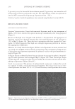
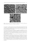
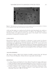
JOURNAL OF COSMETIC SCIENCE 228 mode at a magnifi cation ×1000. Immersion oil was deposited between the objective lens and the upper glass lamella. Coated and noncoated liposomes were observed in parallel using freeze-fracture electron microscopy. About 1 μl of liposome suspension was placed on a copper base. The sample was rapidly frozen with a cryoprotectant (glycerol) in liquid nitrogen (-196°C) cooled propane. After storage in liquid nitrogen, samples were placed on the support immersed in liquid nitrogen which was inserted into the cryoscouring device (BAL-TEC BAF 060 (Nanterre, France)) and then cooled to -150°C. When a vacuum of 10-7 torr was reached, samples were fractured using a scalpel blade cooled to -150°C. The fracture surface was shadowed by a spray of platinum, and a layer of 4 nm was fi lled in one direction at an angle of 45°C to reveal the relief and surface structures. The thin layer was consolidated with vertically sprayed carbon, which is transparent to electrons. A quartz oscillating measuring device measured the fi nal thickness it was about 30 nm. The replicas were cleaned with a mix- ture of solvents and/or sulfochromic acid, rinsed with distilled water, and examined under a transmission electron microscope (FEI CM120, Philips, Cambridge, UK). Vesicle size measurement. The empty noncoated liposomes size was measured by dynamic light scattering (DLS) using a Malvern Zetasizer Nano ZS (Malvern Instruments, Worces- ter, UK). The samples were diluted (1:400) with ultrafi ltrated distilled water. Droplets sizes were obtained from the correlation function calculated by the dispersion technology software using various algorithms. The apparatus is equipped with a 4 mW He/Ne laser, emitting 633 nm, measurement cell, photomultiplier, and correlator. The refractive in- dex and absorbance were set at 1.47 and 0.01 respectively at 25°C. The measurements were performed in fi ve replicates (19). DLS is not adapted to polydisperse suspensions and important size vesicles. Therefore coated liposomes sizes were determined using static light scattering. The size distribution of empty coated liposomes and MCCL was determined using static light scattering (Beckman Coulter LS230 instrument, Brea, CA) according to Fraunhofer diffraction theory. By applying the polarization intensity differential scattering, this ap- paratus can detect particles from 0.04 to 2000 μm using up to 116 size classes and a he- lium neon laser with a wavelength of 750 nm. Empty coated liposomes and MCCL suspensions were dispersed in distilled water under agitation into the measuring cell to be in the optimal obscuration range (8%–20%). Par- ticle size distribution was expressed in volume units. STABILITY EVALUATION Resistance to surfactants and electrolytes. Since most cosmetic formulations are emulsions, the improvement of liposome stability against surfactants is a very important aspect for their use in such cosmetic formulations. Empty coated and noncoated liposomes were incubated for 30 days at 25°C with different percentages of ionic surfactants (SDS, SLES), nonionic surfactants (Triton X-100, Poly- sorbate 20, polyglyceryl-10-laurate, Behenyl alcohol ethoxylate), and salts (NaCl, MgCl2). As high as 90% of liposome suspensions were mixed with 10% of surfactant solutions at different concentrations. Surfactant solutions were prepared in distilled water at different

NEW RESISTANT LIPOSOME COATED WITH POLYSACCHARIDE FILM FOR COSMETIC APPLICATION 229 concentrations from 1% to 10%. Regarding the salts resistance test, 70% of coated and noncoated liposome suspensions were mixed with 30% of various salt solutions. The fi nal concentrations of salts in liposome suspensions were set at 5%, 10%, 15%, 20%, 25%, and 30%. All fractions were expressed as w/w. During a period of 30 days, daily optical microscope observations were performed at a magnifi cation ×1000 at 25°C. We believe that the liposome suspension is unstable from the fi rst signs of destabilization, such as aggregation and destruction of the phospholipid bilayer. PERMEABILITY EVALUATION Magnesium chloride release from coated liposomes. Twelve percent of MgCl2 was entrapped in coated liposomes. The permeability of coated liposome membrane to MgCl2 was evalu- ated in acrylates/C10-30 alkyl acrylate cross-polymer hydrogels (0.8%, w/w) neutralized with sodium hydroxide. Being sensitive to electrolytes, the viscosity of hydrogels de- creases by increasing the concentration of MgCl2. The release of MgCl2 from coated lipo- somes was then evaluated by measuring the viscosity evolution of hydrogels containing 2% (w/w) of entrapped MgCl2 in coated liposomes (MCCL) and an equivalent amount of free MgCl2 (0.24% w/w). The viscosity measurement was performed using a rheometer apparatus (Rheomat RM 200, Lamy Rheology, Champagne au Mont d’Or, France). The effect of coated liposome dilution (1/2, 1/10) on the release of MgCl2 was carried out. We supposed that dilution of the external medium of coated liposomes (distilled water) could increase the diffusion of MgCl2 from their internal core. EVALUATION OF THE DELIVERY EFFICACY OF COATED LIPOSOMES In order to bring out the delivery effi cacy of coated liposomes, the latter were used to improve the detoxifi cation activity of MgCl2. Human skin explants were used to evaluate the effect of entrapped and free MgCl2 on P-gp transporter activity. Human normal skin explants were obtained from a piece of surgical resection of 37-year-old subjects. Hydro- gel composed of 1% (w/w) of acrylates/C10-30 alkyl acrylate cross-polymer neutralized with sodium hydroxide, 0.5% (w/w) of xanthan gum and preserved with 0.8% (w/w) of parabens and phenoxyethanol’s mixture was used. Deionized water was added to com- plete the formulation (100% w/w). Entrapped and free MgCl2 was incorporated into the hydrogel at 3% (i.e., 0.36% of MgCl2) and 0.36%, respectively. Hydrogel without coated liposomes and MgCl2 was used as placebo. A mixture of ascorbic acid (100 μg/ml) and glutathione (100 μg/ml) was tested as a positive control for the evaluation of P-gp activ- ity. Skin explants were incubated during 24 h at 37°C in contact with positive controls, entrapped MgCl2, nonentrapped MgCl2, and hydrogel as placebo. All formulations were applied to the skin explants’ surface except the reference product, which was directly diluted in the culture medium. At the end of the incubation period, skin explants were rinsed with a PBS and stored at -196°C. The assay was performed in triplicate. Proteins assay. At the end of the incubation period, proteins were quantifi ed in cell lysates by a spectrocolorimetric method using bicinchoninic acid assay (20).
Purchased for the exclusive use of nofirst nolast (unknown)
From: SCC Media Library & Resource Center (library.scconline.org)