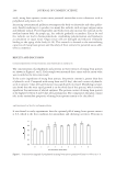
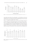
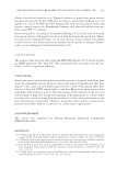
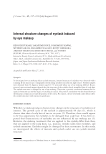
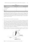
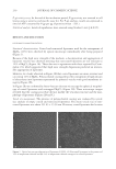
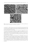
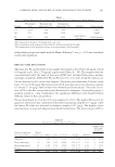
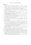
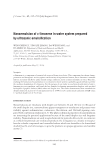
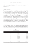
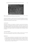
JOURNAL OF COSMETIC SCIENCE 230 P-gp activity assay. At the end of the incubation period, P-gp activity was assessed in cell lysates using a sensitive and specifi c assay kit. For P-gp analysis, results are expressed as nmol of ATP consumed by P-gp per μg of proteins (mean ± S.D.). Statistical analysis. Levels of signifi cance were assessed using Student t-test (p ≤ 0.05). RESULTS AND DISCUSSION LIPOSOMES CHARACTERIZATION Structural characterization. Coated and noncoated liposomes used for the entrapment of MgCl2 (12%) were observed by optical microscope immediately after being prepared (Figure 1). Because of the high ionic strength of the medium, a decomposition and aggregation of liposome vesicles was observed attesting that noncoated liposomes are not resistant to 12% of MgCl2 (Figure 1A). These data are in agreement with those reported by Crom- melin (21), which supported that high ionic strength dispersions predicted an irrevers- ible aggregation of liposomes. However, we clearly observed in Figure 1B that coated liposomes are more resistant and can entrap 12% of MgCl2. Polysaccharide coating allowed the entrapment of high amount of electrolytes into liposomes represented by spherical vesicles with good membrane in- tegrity (Figure 1B). In Figure 2B, we confi rmed by freeze-fracture electron microscopy the spherical morphol- ogy of coated liposomes with entrapped MgCl2 (Figure 2B). These microscope images revealed that the coating procedure did not modify the vesicular structure and the mor- phology of liposomes (Figures 2B and C). Vesicle size measurement. The presence of polysaccharide coating was evaluated by vesicle size analysis of empty coated and noncoated liposomes. The mean vesicle size of non- coated liposomes was about 251.03 ± 15.56 nm. However, coated liposomes had a mean Figure 1. Optical microscope observation of liposomes (×1000): (A) Noncoated liposomes in the presence of 12% of magnesium chloride and (B) 12% of magnesium chloride entrapped in coated liposomes.

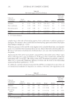
NEW RESISTANT LIPOSOME COATED WITH POLYSACCHARIDE FILM FOR COSMETIC APPLICATION 231 size of 514.00 ± 79.00 nm and thus are two times larger than noncoated liposomes. This size difference could be explained by the presence of a polysaccharide fi lm (11–16). The stearic acid from Stearoyl Inulin would be anchored inside the liposomal membrane with the inulin surrounding the outer bilayer. On the other hand, the entrapment of MgCl2 into coated liposomes (MCCL) affected their size and their size distribution. In the presence of MgCl2, coated liposome size was in- creased without impacting their morphology (Figures 2A, B, and C). The mean size of MCCL was about 1.37 μm, and 90% of vesicles had a size inferior to 3.14 μm. The in- crease in liposome size might be linked to the swelling of polysaccharide fi lm in contact with high amounts of salts (22). Beyond size parameter modifi cation, the introduction of a hydrophobic chain into lipo- some membrane may affect the physicochemical properties of liposomes. According to Carafa et al. (11), coating by hydrophobic chain grafted polysaccharides decreased transi- tion temperature of liposome phospholipids compared to nongrafted polysaccharides. We also supposed that hydrogen bonds and hydrophobic interactions between carbon chain of phospholipids and stearic acid from hydrophobized polysaccharides could limit the release of the encapsulated molecules by forming a very tight embedment in the membrane. Figure 2. Freeze-fracture electron microscopy (×41,100): (A) Empty noncoated liposomes, (B) Magnesium chloride entrapped in coated liposomes, and (C) Empty coated liposome.
Purchased for the exclusive use of nofirst nolast (unknown)
From: SCC Media Library & Resource Center (library.scconline.org)