SACRAN PROTECTS SKIN AGAINST POLLUTANTS 19 MATERIALS AND METHODS MATERIALS Sacran was extracted from the algae Aphanothece sacrum (Suizenji-nori) and was purifi ed (19). Bio-sodium hyaluronate (MMW), as a general HA, was obtained from SK Bioland (Chungnam, Cheonan, South Korea), and 1,3-butylene glycol was obtained from Kokyu Alcohol Kogyo Co. (Tokyo, Japan). Dulbecco’s modifi ed Eagle medium (DMEM) and Hanks’ Balanced Salt solution with Ca2+ and Mg2+ (HBSS) were obtained from Nissui Pharmacy (Tokyo, Japan), and fetal bovine serum (FBS) was obtained from Invitrogen (Carlsbad, CA). The BCA Protein Assay Reagent kit was purchased from Pierce Chemical Co. (Rockford, IL). LabCyte EPI-MODEL reconstructed human epidermal equivalents (RHEEs) at 12 d, and their assay medium were obtained from Japan Tissue Engineering (Aichi, Japan). 6-[6-(Biotinylamino)hexanoylamino]hexanoylhydrazine (biotin-AC5- hydrazide) and Hoechst 33342 solution were obtained from Dojindo Laboratories (Kumamoto, Japan). DyLight650-labeled streptavidin was obtained from Thermo Fisher Scientifi c Inc. (Waltham, MA). Acrolein monomer, NBD-hydrazine (4-hydrazino-7-nitrobenzofurazan- hydrazine), and 4-(4,6-dimethoxy-1,3,5-triazin-2-yl)-4-methylmorpholinium chloride were purchased from Tokyo Chemical Industry Co. (Tokyo, Japan). Fluorescein- 5-thiosemicarbazide (FTSC), 2′,7′-dichlorodihydrofl uorescein diacetate (H2DCFDA), and BaP were purchased from Sigma-Aldrich (St. Louis, MO). Dimethyl sulfoxide was purchased from Nacalai Tesque (Kyoto, Japan). 2-Mercapto-ethanol (2-ME) and SYBR® Green Real-Time polymerase chain reaction (PCR) Master Mix for real-time PCR analy- sis were obtained from Thermo Fisher Scientifi c Inc. (Kanagawa, Japan). RLT buffer and the RNeasy Mini Kit were purchased from Qiagen (Hilden, Germany). SACRAN AQUEOUS SOLUTION A 0.05% (w/v) sacran aqueous solution was used for experiments as noted in the text. As a representative anionic polysaccharide that is commonly formulated in skin care products, we used a 0.05% (w/v) HA aqueous solution. BIOTIN CONJUGATION TO POLYSACCHARIDES Polysaccharides were reacted with 2 mg/mL biotin-(AC5)2-hydrazide in the presence of 20 mg/mL 4-(4,6-dimethoxy-1,3,5-triazin-2-yl)-4-methylmorpholinium chloride and pyridine for 5 h. Biotin-conjugated polysaccharides were purifi ed by dialysis against de- ionized water. PENETRATION OF POLYSACCHARIDES INTO RHEEs The penetration of biotin-conjugated polysaccharides through RHEEs was measured by quantifi cation of biotin-conjugated polysaccharides in the culture medium using an Enzyme- linked immunosorbent assay (ELISA) and fl uorescence histology. RHEEs were topically treated with biotin-conjugated polysaccharides, and then were cultured for 24 h at 37°C.

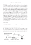
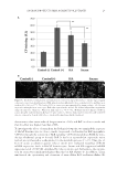
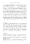
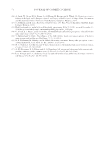
JOURNAL OF COSMETIC SCIENCE 20 Each RHEE was separated into two pieces with a scalpel. Frozen thin sections of one piece of each RHEE embedded in optimal cutting temperature compound were prepared using a cryomicrotome, and the localization of biotin-conjugated polysaccharides in RHEEs was visualized by staining with DyLight650-labeled streptavidin. Nuclei were stained with Hoechst33342. Fluorescence images were taken with a Floid Cell Imaging Station (Thermo Fisher Scientifi c Inc.). The other piece of each RHEE was used to extract biotin-conjugated polysaccharides following homogenization in 500 μL phosphate-buffered saline without Ca2+ and Mg2+ [PBS(-)] at 2,700 rpm for 10 min with a μT-12 bead crusher (Taitec Corp., Saitama, Japan). Biotin-conjugated polysaccharides remaining in the RHEEs or penetrating into the culture medium through the RHEEs were quantifi ed using an ELISA method. Briefl y, the RHEE extract or culture medium was incubated with horseradish peroxidase-conjugated streptavidin (1:1,000) for 1 h at 37°C. The mixed solution was placed in wells of ELISA plates (MS-8596F, Sumitomo Bakelite, Tokyo, Japan) coated with biotin-conjugated bovine serum albmin (BSA), and then incubated for 1 h at 37°C. Each well was washed with PBS-T, then 150 μL 2,2′-azinobis (3-ethylbenzothiazoline- 6-sulfonic acid) diammonium salt (ABTS Wako, Osaka, Japan) solution (0.3 mg/mL) in phosphate-citrate buffer (0.1 M, pH 4.0) containing a small amount of H2O2 was added to each well. After 30 min, the absorbance of each well was measured using a microplate reader (Spark 10M TECAN, Männedorf, Switzerland) at 405 nm. Amounts of biotin- conjugated polysaccharides were determined using a calibration curve prepared with biotin as a standard substance. TOBACCO SMOKE Seven Stars® (tar: 14 mg, nicotine 1.2 mg, Japan Tobacco, Tokyo, Japan) was used a source of tobacco smoke. TRAPPING EFFECTS OF POLYSACCHARIDES AGAINST TOBACCO SMOKE The trapping effects of polysaccharides against tobacco smoke were examined by measuring ACs and BaP in PBS diffused with tobacco smoke using fl uorescence methods (Figure 1). The smoke obtained from burning one piece of tobacco was introduced into PBS stirred with a magnetic stirrer passing through membrane fi lters (10 μm JH, Merck Millipore, Burlington, MA) that had been treated with or without polysaccharide by soaking in 1 mL polysaccharide Figure 1. Method for treatment by tobacco smoke to PBS(-).
Purchased for the exclusive use of nofirst nolast (unknown)
From: SCC Media Library & Resource Center (library.scconline.org)