JOURNAL OF COSMETIC SCIENCE 210 DNA DAMAGE Sunlight, primarily the shorter wavelengths in the UV range, are absorbed by the DNA in living skin cells, producing a variety of direct chemical modifi cations as well as modi- fi cations by reactive molecules secondarily produced by sunlight (recently reviewed in ref. 6). The most common form is the cyclobutane pyrimidine dimer (CPD, fusion of adjacent DNA bases) followed by the (6-4) photoproduct (6-4PP, also a fusion of DNA bases) and oxidation of the DNA base guanine producing 8-oxo-guanine (8oGua). On a macroscopic level, the occurrence of CPDs is randomly distributed along the genome at dipyrimidine sites, but closer examination has revealed that nucleosomes infl uence DNA damage for- mation and repair (7), and hotspots adjacent to certain transcription-binding sites prefer- entially accumulate DNA damage.(8) Telomeres, the special tips of chromosomes, are especially susceptible to damage due to the high density of dipyrimidines (9). Long wave ultraviolet A (UVA) (UVA1 340–400 nm) produces CPD with a predilection for the basal epidermis where the actively dividing stem cells reside, and thus broad band photopro- tection is important for reducing the DNA damage burden (10) . The predominant mutation in keratinocyte tumors is the ultraviolet radiation (UVR) signature mutation (11,12), and the CPD is of particular trepidation for skin health, as not only does its formation triggers erythema and the sunburn reaction (13) but also immune suppression that allows the outgrowth of skin tumors (14). Sunscreens are less effi cient in preventing immunosuppression than blocking erythema, perhaps because only small amounts of the CPD are able to initiate it (15) . Recent research highlights the role of UVA in melanoma development, including DNA damage in melanocytes and inhibition of DNA repair (16). Of particular interest is a study showing that melanin by-products absorb UVA and continue to form CPDs even in the absence of UVA (17). Thus, pigmentation not only protects skin from UV dam- age but may, in some cases, also foster it. Pigmentation occurs after UV-induced stimulation of α-melanin stimulating hormone production which then binds to melano- cortin 1 receptor, promoting tyrosinase activity and melanin formation, and may also stimulate DNA repair (16). On the other hand, another melanocyte-specifi c transcription regulator was shown to turn up pigmentation and turn down DNA repair, and vice versa, in a counterbalancing system (18). DNA REPAIR The broad outlines of nucleotide excision repair (NER) of UV-induced DNA damage were recently reviewed (19,20) A complex of proteins, many also involved in gene tran- scription, identify distortions in DNA produced by photoproducts, and phosphorylation of the xeroderma pigmentosum group C protein within this complex recruits the rest of the NER proteins to the damaged site (21). A length of single-stranded DNA containing the lesion is excised, and the opposite intact strand serves as a template to fi ll in the gap. When DNA replication uses a damaged template, an error-prone polymerase enables replication across the lesion, at the cost of somatic mutations, but with an overall reduc- tion in skin cancer incidence (22). Whereas the entire genome is surveilled for damage by a global repair system, (23) a special system of transcription-coupled repair focuses repair complexes at transcription sites (24,25). This interactive relationship between NER and gene expression was recently

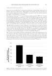
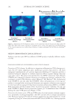
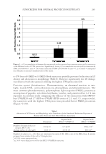
IMPORTANCE OF DNA REPAIR 211 reviewed (26). The physical relationship between transcription factor transcription factor IIH and the NER factor xeroderma pigmentosum group A was visualized, which explains how transcription factors are recruited to NER complexes (27). The complexity of repair- ing DNA bound in nucleosomes and chromatin (7) is solved by the binding of UV-DNA damage-binding protein to UV-damaged nucleosomes and the shifting of nucleosome structure to expose DNA damage (28). An added complexity is the ability of cellular signaling pathways, such as those regulated by cytokines, to modify NER according to the state of the cell, organ, or body (29). Circadian rhythm and the molecular clock affect DNA repair and related responses such as pigmen- tation (30). One consequence of these DNA repair oversight functions is that low, chronic doses increase expression of DNA repair proteins and result in increased repair of CPD but not (6–4) PP (31). Recruitment of DNA repair complexes to damaged sites starts in 1 h and peaks at 6 h (32). (6–4) PP are repaired much faster than the CPD because of their more effi cient recognition and base fl ipping by XPC-Rad4 protein complex (33). Most cellular responses peak at around 6 h while infl ammation, and antigen-specifi c immune suppression signals crest at 24 h. Overall, the half-life of CPDs in the human skin is about 11 h when the system is within its capacity (35), but it becomes saturated just at UV doses that produce a sunburn (36). ENHANCING DNA REPAIR The DNA repair capacity of the skin can be enhanced by delivering DNA repair enzymes. The fi rst patent for a commercial method, using phospholipid liposomes encapsulating enzymes to deliver to skin, was granted in 1991 (37) and enables the delivery of a number of enzymes from a variety of microbial sources, including photolyase from Anacystis nidu- lans, UV endonuclease from Micrococcus luteus, bacteriophage T4 endonuclease V, and 8oGua glycosylase 1 from Arabidopsis thaliana. Recently, others have encapsulated the UV DNA damage endonuclease from yeast and the pyrimidine dimer glycosylase from Paramecium bursaria chlorella virus-1 (38). In each case, the liposome–enzyme composition increased the repair of UV-induced DNA damage. Today, more than 75 skincare products are available that contain DNA repair enzymes, and dozens of clinical studies have reported prevention and enhanced regression of actinic keratosis, nonmelanoma skin cancers, and photoaging (reviewed in ref. 39,40). Indeed, adding DNA repair enzymes to sunscreens provides additive protection (41). The benefi ts appear in a few months, suggesting that enhanced DNA repair reduces short-term cancer- promoting signaling and long-term mutagenic events. Another approach uses nicotinamide (vitamin B3) to overcome UV-induced energy de- pletion and subsequent inhibition of DNA repair (42), and this also reduces the erythe- mal response to a given dose of UV. Studies show that daily ingestion of nicotinamide reduces the number of nonmelanoma skin cancers over a 1-year period in Caucasian skin (42). An intriguing advance was the demonstration that secreted proteins from amnion- derived multipotent progenitor cells applied topically on the human skin immediately after UV irradiation reduced erythema, increased XPA DNA repair protein, and decreased DNA damage (43). Similarly, extracellular vesicles derived from human adipose-derived stem cells, which contain a mixture of miRNAs and proteins, mitigated many effects of UVB irradiation (44).
Purchased for the exclusive use of nofirst nolast (unknown)
From: SCC Media Library & Resource Center (library.scconline.org)