JOURNAL OF COSMETIC SCIENCE 218 acute endpoint to determine personal UVR sensitivity is erythema (i.e., sunburn), in particular the minimal erythema dose (MED). This is the lowest physical UVR dose (J/cm2) that gives just perceptible skin redness about 24 h after exposure. Melanocytes are dendritic cells that reside in the basal layer of the epidermis. They syn- thesize melanin that is transferred to epidermal keratinocytes that are the majority cell type. This process is enhanced (i.e., tanning) when the skin is exposed to UVR. Melanin is mostly concentrated in the lower epidermis and also presents as caps over nuclei of basal layer keratinocytes. Several studies have attempted to enhance the tanning response with a view to improving photoprotection. Some have used sunscreen formulations that include 5-methoxypsoralen (5-MOP), which is a natural photosensitizing ingredient in bergamot (Citrus bergamia) oil (6). However, this approach is not viable because 5-MOP is a photocarcinogen (7). An important factor in tanning is the binding of α-melanocyte–stimulating hormone (α-MSH) to the melanocortin 1 receptor on melanocytes. Another approach has been to generate synthetic analogues of α-MSH to enhance tanning (8). The re is a melanin gradient with terrestrial UVB in indigenous populations. Melanin decreased as humans in prehistory dispersed away from tropical latitudes, and one driving factor for this loss is thought to be the need to maintain adequate vitamin D synthesis (9,10). Mass migration and travel in relatively recent history has meant people’s skin color no longer matches the solar conditions under which it evolved. The need for photo- protection in normal healthy people depends on the FST, the intensity (irradiance) of the sun, and the time spent outdoors with uncovered skin. The World Health Organization has promoted the ultraviolet index (UVI) as means of estimating the sunburning capacity of solar UVR at any given location. The categories are low (1–2), moderate (3–5), high (6–7), very high (8–10), and extreme (11+). Photoprotection is recommended for light skin when the UVI reaches 3, but this has raised some controversy (11). SUN SCREENS Sun screens are topical formulations that contain synthetic organic UVR fi lters and/or mineral and/or organic nanoparticles (12). They work by absorbing solar UVR and also by some scattering in the case of pigments. Ideally, they should remain on the surface of the skin, but there is increasing evidence that they can penetrate into the systemic circulation because they or their metabolites can be detected in blood and urine (13). Sunscreen effi cacy is measured by their ability to prevent erythema ~24 h after exposure to solar simulated radiation (SSR). The index for this is the sun protection factor (SPF) when the test prod- uct is applied at 2 mg/cm2 skin. Essentially, this is a ratio of the MED with and without sunscreen application (14). SSR does not contain visible light (400–700 nm), and sun- screens are not designed to protect in this region. It has been suggested the laboratory- determined SPF may underestimate protection against erythema because of not taking visible radiation into account (15). COM PARISONS OF PHOTOPROTECTION BY MELANIN AND SUNSCREENS Mel anin, especially constitutive melanin, has the advantage of being always present. Fur- thermore, its presence increases after exposure to sunlight. Sunscreens, on the other hand,

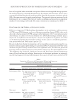
SKIN PHOTOPROTECTION BY PIGMENTATION AND SUNSCREENS 219 have to be applied before intended sun exposure behavior and reapplied during exposure. No study has directly compared photoprotection by melanin and sunscreens. In fact, this is conceptually diffi cult because the results would be biased by the amount of melanin and the SPF of the sunscreen and its application thickness. The approach taken as mentioned in the following text is to compare the results of studies on melanin and sunscreens on important markers of solar damage and benefi t. The main conclusions are summarized in Table I. DNA DAMAGE, ERYTHEMA, AND SKIN CANCER DNA is an important UVR-absorbing chromophore in the epidermis, and the presence of UVR causes DNA damage, such as cyclobutane pyrimidine dimers (CPDs), that can be detected in human skin after UVR exposure in the laboratory (16,17) and in skin or urine after holidays in adults (18,19) and children (20). DNA photodamage probably plays a major role in erythema and certainly has a major role in photocarcinogenesis (21). This can be demonstrated by the very high incidence of skin cancer in xeroderma pigmento- sum patients who lack DNA repair capacity (22). Rec ent studies have shown the importance of location when assessing protection by con- stitutive melanin against photodamage to epidermal DNA. Comparisons of FST II versus VI show that protection by melanin varies with epidermal zone, such that the protection factor by melanin in the basal layer is about 60 but only fi ve in the upper epidermis (23). This difference has biological signifi cance because the basal layer contains keratinocyte stem cells and melanocytes, whereas keratinocytes in the upper epidermis are approach- ing the fi nal stages of terminal differentiation. Skin cancer is much more common in FST Table I Comparis on of Protection by Pigmentation (Melanin) and Sunscreens for Adverse and Benefi cial Effects of Solar UVR Endpoint Type and level of protection Constitutive pigmentation Facultative pigmentation Sunscreen DNA damage (CPD) Very high protection factor of ~60 in basal layer of the epidermis when comparing FST VI with II (23). Modest, with protection factors in the region of 2–4 (29). Depends on the SPF and application thickness (39). May be very high with high SPF sunscreen (40). Erythema About 6- to 8-fold when comparing FST VI with II (25,26). Modest, with protection factors in the region of 2–4 (27–29). Depends on the SPF in laboratory studies but no data on the “real-life” SPF. Very effective when used correctly on holiday (18). Skin cancer High (5). Unknown Low (42–44). Vitamin D synthesis Low, with an estimated inhibitory factor of 1.3 when comparing FST VI with II (51). Unknown Low but very few data from intervention studies (52–54). Studies needed on high SPF intervention (55).
Purchased for the exclusive use of nofirst nolast (unknown)
From: SCC Media Library & Resource Center (library.scconline.org)