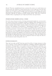
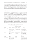
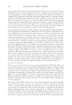
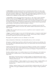
SKIN PHOTOPROTECTION BY PIGMENTATION AND SUNSCREENS 219 have to be applied before intended sun exposure behavior and reapplied during exposure. No study has directly compared photoprotection by melanin and sunscreens. In fact, this is conceptually diffi cult because the results would be biased by the amount of melanin and the SPF of the sunscreen and its application thickness. The approach taken as mentioned in the following text is to compare the results of studies on melanin and sunscreens on important markers of solar damage and benefi t. The main conclusions are summarized in Table I. DNA DAMAGE, ERYTHEMA, AND SKIN CANCER DNA is an important UVR-absorbing chromophore in the epidermis, and the presence of UVR causes DNA damage, such as cyclobutane pyrimidine dimers (CPDs), that can be detected in human skin after UVR exposure in the laboratory (16,17) and in skin or urine after holidays in adults (18,19) and children (20). DNA photodamage probably plays a major role in erythema and certainly has a major role in photocarcinogenesis (21). This can be demonstrated by the very high incidence of skin cancer in xeroderma pigmento- sum patients who lack DNA repair capacity (22). Rec ent studies have shown the importance of location when assessing protection by con- stitutive melanin against photodamage to epidermal DNA. Comparisons of FST II versus VI show that protection by melanin varies with epidermal zone, such that the protection factor by melanin in the basal layer is about 60 but only fi ve in the upper epidermis (23). This difference has biological signifi cance because the basal layer contains keratinocyte stem cells and melanocytes, whereas keratinocytes in the upper epidermis are approach- ing the fi nal stages of terminal differentiation. Skin cancer is much more common in FST Table I Comparis on of Protection by Pigmentation (Melanin) and Sunscreens for Adverse and Benefi cial Effects of Solar UVR Endpoint Type and level of protection Constitutive pigmentation Facultative pigmentation Sunscreen DNA damage (CPD) Very high protection factor of ~60 in basal layer of the epidermis when comparing FST VI with II (23). Modest, with protection factors in the region of 2–4 (29). Depends on the SPF and application thickness (39). May be very high with high SPF sunscreen (40). Erythema About 6- to 8-fold when comparing FST VI with II (25,26). Modest, with protection factors in the region of 2–4 (27–29). Depends on the SPF in laboratory studies but no data on the “real-life” SPF. Very effective when used correctly on holiday (18). Skin cancer High (5). Unknown Low (42–44). Vitamin D synthesis Low, with an estimated inhibitory factor of 1.3 when comparing FST VI with II (51). Unknown Low but very few data from intervention studies (52–54). Studies needed on high SPF intervention (55).

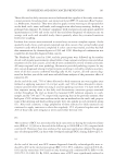
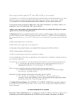
JOURNAL OF COSMETIC SCIENCE 220 I/II versus V/VI, and interestingly, the difference in the incidence of basal cell carcinoma (BCC), the most common type of skin cancer, is about 60 (23). The re is an inverse relationship between the increasing FST and the MED on habitually sun-protected skin. However, there is considerable overlap of the MED between FST I and IV. The difference in the MED between FST II and IV on sun-protected skin is about two, which is very modest in SPF terms (24). There have been fewer studies with FST V and VI, but comparisons between I/II and VI show a protection factor of melanin against erythema of about 6–8 (25,26). This is about the same as the protection factor against DNA photodamage for the whole epidermis (23). Stu dies to determine the protective properties of facultative tanning in FST I–IV have shown this to be modest against DNA damage and erythema. Field and laboratory stud- ies have shown protective factors to be in the region of 2–3 (27–29). Other factors such as stratum corneum thickening as a result of solar exposure may be important, but they are poorly understood (30). It is relatively simple to determine the SPF under very controlled laboratory conditions, one of which is the application of the sunscreen at 2 mg/cm2 on skin (14). In practice, people apply very much less with a commensurate typically linear reduction in the SPF (12). For example, one study on Danes on holiday in Egypt showed an average application thickness of 0.79 mg/cm2 (31). This can result in overestimation of protection and, there- fore, overexposure and sunburn (32,33). In general, people do not apply sunscreen very well (34). Correct application of a sunscreen can prevent erythema during a week’s sun holiday with maximal UVI of 9 (18). Sun screens are effective at preventing epidermal CPDs (35), which would be expected because erythema and epidermal CPDs have similar action spectra in the solar UVR range (36). CPDs can be caused by suberythemal exposure, and such DNA damage (as well as erythema) accu- mulates with such exposure daily because epidermal CPDs have a half-life of 33 h (i.e., slow repair) (37). Daily application for 11 d of a low SPF sunscreen, before suberythemal SSR expo- sure, was shown to be effective at reducing cumulative CPDs and erythema (38). A more re- cent study, which detected CPDs by immunostaining and quantitative high pressure liquid chromatography with tandem mass spectrometry, has shown that an SPF 50+ sunscreen was very effective at preventing DNA damage by very high doses of acute and repeated (fi ve con- secutive days) SSR exposure (39). This study also showed that DNA protection, in acute and repeat SSR exposure cases, was dependent on sunscreen application thickness. Signifi cant pro- tection was observed even with application at 0.75 mg/cm2 (equivalent to an SPF of 21), typical of consumer use. Studies have not been designed to determine a DNA protection factor (DNA-PF). A new approach has been recently reported in which an SPF 50+ sunscreen had a DNA-PF of 98.2 [95% confi dence interval (CI) of 51.6–187.2] (40). The re is considerable evidence that the CPD plays a major role in skin cancer, especially keratinocyte cancers (21). Thus, they would be expected to have a benefi cial effect against human skin cancer as has been reported in studies on mouse models (41). Studies show that sunscreen use can inhibit skin cancer (42). The best prospective evidence is from long-term intervention studies in Nambour, Queensland, Australia (discussed in the ar- ticle by A. Green in the same issue). The most robust conclusion from these studies is that a 4.5-y randomized controlled intervention with an SPF 16 product signifi cantly reduced the incidence of melanoma (43) and squamous cell carcinoma (SCC), but not BCC (44). This reduction is very important clinically, but less so numerically for example, the rate
Purchased for the exclusive use of nofirst nolast (unknown)
From: SCC Media Library & Resource Center (library.scconline.org)