131 ImprovedPROVED AVB PhotostabilityOTOSTABILITY UsingING NLCs
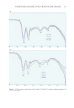
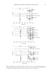
AVB:lipid-based excipients binary mixtures (1:1 m/m) were analyzed at wavelengths of
4,000–600 cm−1 with a resolution of 4 cm−1, using attenuated total reflection (ATR).
Around 2 ± 1 mg were used to evaluate the solid excipients and 20 uL (about a drop) of
the liquids. Each sample was subjected to 64 scans to obtain the spectra.
DEVELOPMENT AND IN VITRO CHARACTERIZATION OF THE
NANOSTRUCTURED LIPID CARRIERS
NANOSTRUCTURED LIPID CARRIER OBTAINING
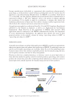
After the evaluation of AVB compatibility with NLC excipients, the formulations were
obtained by the phase inversion method previously described by Shinoda et al.25 Water
and oil phase components were weighed separately. The lipid, surfactant, and AVB were
weighed and then added to the same container. For the water phase, water was weighed in a
separate container. Both phases were heated to a temperature of 80–90°C and, subsequently,
the water phase was poured into the oil phase under mechanical stirring at 400 rpm and
then left to stabilize for 10 minutes. The formulation (Table 1) was submitted to high-
pressure homogenization at 400 bar in a LV1 Microfluidizer (Microfluidics Corporation,
Massachusetts, USA). 0.5% AVB was added to the oil phase.
PHYSICOCHEMICAL CHARACTERIZATION OF NLCS
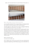
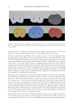
The formulations were evaluated regarding their macroscopic aspect, average particle
diameter, polydispersity index (PdI), zeta potential, and encapsulation efficiency (EE%).
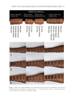
Color, homogeneity, presence, or absence of coalescence or precipitate were visually
evaluated. The average diameter and PdI were evaluated by dynamic light scattering using
a ZetaSizer Nano S (Malvern Instruments Ltd., Worcestershire, UK) using a 1/40 dilution
in ultrapure water. Zeta potential was evaluated by electrophoretic mobility in a ZetaPlus
equipment (Brookhaven, New York, USA) using a 1/40 dilution in ultrapure water. The
EE% was evaluated by the indirect method, in which 1 mL of the NLC dispersion was
centrifuged (SIGMA 3-18K Centrifuge®, SciQuip, Shrewsbury, UK) at 12,000 rpm for 20
minutes. The collected supernatant was diluted with ACN, homogenized, and quantified
by HPLC. Drug EE was calculated according to Eq. 1.
EE (%)Amount of AVB in the NLC
Amount of AVB add in the formulation. 1000 =× (Eq. 1)
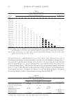
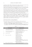
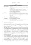
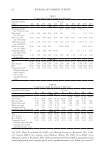
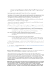
Table I
Percentual Composition of NLC Formulations Evaluated for the
Encapsulation of AVB
Composition F1 F2 F3
Carnauba wax 3.5 3.5 2.5
Isopropyl myristate 1.5 1.5 2.5
Sorbitane trioleate (Span 85) 1.25 1.75 1.89
Polysorbate 80 (Tween 80) 3.75 5.25 5.11
Water qs 100 qs 100 qs 100
AVB:lipid-based excipients binary mixtures (1:1 m/m) were analyzed at wavelengths of
4,000–600 cm−1 with a resolution of 4 cm−1, using attenuated total reflection (ATR).
Around 2 ± 1 mg were used to evaluate the solid excipients and 20 uL (about a drop) of
the liquids. Each sample was subjected to 64 scans to obtain the spectra.
DEVELOPMENT AND IN VITRO CHARACTERIZATION OF THE
NANOSTRUCTURED LIPID CARRIERS
NANOSTRUCTURED LIPID CARRIER OBTAINING
After the evaluation of AVB compatibility with NLC excipients, the formulations were
obtained by the phase inversion method previously described by Shinoda et al.25 Water
and oil phase components were weighed separately. The lipid, surfactant, and AVB were
weighed and then added to the same container. For the water phase, water was weighed in a
separate container. Both phases were heated to a temperature of 80–90°C and, subsequently,
the water phase was poured into the oil phase under mechanical stirring at 400 rpm and
then left to stabilize for 10 minutes. The formulation (Table 1) was submitted to high-
pressure homogenization at 400 bar in a LV1 Microfluidizer (Microfluidics Corporation,
Massachusetts, USA). 0.5% AVB was added to the oil phase.
PHYSICOCHEMICAL CHARACTERIZATION OF NLCS
The formulations were evaluated regarding their macroscopic aspect, average particle
diameter, polydispersity index (PdI), zeta potential, and encapsulation efficiency (EE%).
Color, homogeneity, presence, or absence of coalescence or precipitate were visually
evaluated. The average diameter and PdI were evaluated by dynamic light scattering using
a ZetaSizer Nano S (Malvern Instruments Ltd., Worcestershire, UK) using a 1/40 dilution
in ultrapure water. Zeta potential was evaluated by electrophoretic mobility in a ZetaPlus
equipment (Brookhaven, New York, USA) using a 1/40 dilution in ultrapure water. The
EE% was evaluated by the indirect method, in which 1 mL of the NLC dispersion was
centrifuged (SIGMA 3-18K Centrifuge®, SciQuip, Shrewsbury, UK) at 12,000 rpm for 20
minutes. The collected supernatant was diluted with ACN, homogenized, and quantified
by HPLC. Drug EE was calculated according to Eq. 1.
EE (%)Amount of AVB in the NLC
Amount of AVB add in the formulation. 1000 =× (Eq. 1)
Table I
Percentual Composition of NLC Formulations Evaluated for the
Encapsulation of AVB
Composition F1 F2 F3
Carnauba wax 3.5 3.5 2.5
Isopropyl myristate 1.5 1.5 2.5
Sorbitane trioleate (Span 85) 1.25 1.75 1.89
Polysorbate 80 (Tween 80) 3.75 5.25 5.11
Water qs 100 qs 100 qs 100