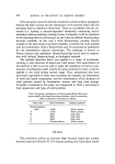
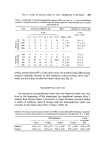
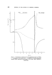
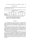
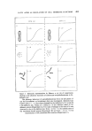
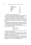
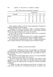
FATTY ACIDS AS REGULATORS OF CELL MEMBRANE FUNCTIONS 491 Chemical analysis Polyunsaturated fatty acid contents of the various lipid fractions of the epidermis are given in Table III. Table III. Determination of essential fatty acids (%) in various lipid fractions of rat epidermis during EFA-deficiency Control rats EFA-deficient rats FFA SE TG PL FFA SE TG PL Linoleic acid 2.9 2.7 6.9 5.2 1.9 1.1 2.3 2.6 Eicosatrienoic acid Trace Trace Trace Trace 0.3 1.7 0.3 3.3 Arachidonic acid 1.1 1.0 Trace 3.6 1.5 2.2 0.1 2.5 FFA, Free fatty acids TG, triglycerides SE, sterol esters PL, phospholipids. Linoleic acid: significant fall in triglycerides and phospholipids. Eicosatrienoic acid: rise in sterol esters and phospholipids. Arachidonic acid: fall in phospholipids (may not be significant). DISCUSSION For the interpretation of the various results, it must be kept in mind that each technique yields information relating to a different kind of cutaneous structure since the phoreographical response originates in the living epidermal cells whereas the water-loss is regulated by the stratum corneum. On the other hand it is obvious that, if SSO is administered orally, any change in the stratum corneum proceeds from a previous change in the living epidermal cells from which this layer is derived. EFA-deficiency primarily affects the living epidermal layers and, secondarily, the kera- tinized cells, after a latent period corresponding to the duration of the keratinization process, i.e. about 18-20 days. This appears clearly when comparing the time-dependent curves for AR and R. Very soon after starting the deficient diet, the phoreographical response undergoes modifications and disappears after 3 weeks, showing the first effects of the deficiency at the level of living epidermal cells. Skin resistance, on the contrary, shows practically no variation for 2 weeks a significant decrease occurred only after about 20 days which delay is consistent with the time required for the keratinization of the first de- ficient cells.

492 JOURNAL OF THE SOCIETY OF COSMETIC CHEMISTS The above hypothesis is supported by the fact that throughout the deficiency-period the water-loss curve and the resistance variation curve have a similar pattern and the decrease in skin resistance corresponds to an increased water-loss. This was to be expected since the rate of water-loss as well as the electrical skin resistance are essentially dependent on the condi- tion of the stratum corneum. The events observed after SSO dosage are indicative of more complex phenomena. Oral ingestion, as expected, causes restoration of the phoreographical response which shows the effect produced on the living cells but, at the same time, there is a considerable increase in resistance and a decrease in water loss. It has been established (5) that any intervention aimed at altering the equilibrium between stratum corneum and germinal cells immediately pro- duces a reaction of the latter in the form of an accelerated keratinization process. After stripping or application of solvent (or detergent solution), skin impedance increases tremendously during the 2 days following the treatment. This is indicative of a keratinization process. Histological studies showed that EFA-deficiency always resulted in a distinct thickening of both the stratum corneum and the malpighian layer (4), also that the mitotic figures were increased. Menton (5) speculated that increased water per- meability of the epidermis might precede or be a stimulus to hyperplasia in an attempt to reduce water-loss by increasing the thickness of the epidermal layer. He observed that EFA-deficient mice kept in a humid atmosphere showed low hyperplasia in the epidermis presumably because of reduced water-loss through the skin. Consequently, it might be thought that for the whole period of time of EFA-deficiency--and owing to the existence of a perturbed stratum cor- neum--considerable mitotic activity is going on. But the division of deficient cells can obviously not restore a normal corneum. This is expressed by an increasing water-loss, decreasing electrical resistance and absence of phoreographical response. With SSO-dosage, however, a normal metabo- lism is restored thereby the germinal cells which had already an increased mitotic rate soon become able to rebuild a normal protective barrier and, thus sheltered, can resume division at the normal rhythm. In other words, the delay observed between the disappearance of the phoreographical response and the increase in water-loss and electrical resistance during the initial stages of EFA-deficiency is due to the existence of a normal mitotic cycle. On the contrary, during the period of restoration following after 7 weeks of EFA-deficiency, a perturbed stratum corneum has been substi- tuted for the former normal one, resulting in an acceleration of the mitotic
Purchased for the exclusive use of nofirst nolast (unknown)
From: SCC Media Library & Resource Center (library.scconline.org)