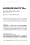
j. Soc. Cosmet. Chem., 39, 121-132 (1988) Improved optical discrimination of skin with polarized light j. PHILP, N. J. CARTER, and C. P. LENN, Environmental SaJ•ty Laboratory, Unilever Research, Colworth House, Sharnbook, Bedford MK44 1LQ, U.K. (J.P., N.J.C.), and Department of Fluid Engineering and Instrumentation, Cranfield Institute of Technology, Cranfield, Bedford MK43 OA, U.K. (C.P.L.). Received July 20, 1987. Synopsis A polarized illumination system is described that optically segregates the skin into two distinct regions, making the assessment of the clinical condition easier. It is possible to highlight, preferentially, the light reflected from the dermis with suppression of light reflected from the surface of the stratum comeurn and vice versa. Polarized reflection spectroscopy and polarized color photography are used to demonstrate the differences between reflected polarized light and light that is depolarized by multiple scattering in the skin. The former shows clear delineation of surface structure whilst the latter highlights the redness or erythemal condition of the skin. INTRODUCTION Visual assessment has always played an important part in evaluating the clinical condi- tion of skin. Trained personnel can estimate the erythemal levels, the degree of rough- ness and dryness, and any other changes that can be visually identified. The major problem with in vivo evaluations is the variety of scales used (1,2) and the discreteness presumed (3). There is also the added difficulty of interpreting the descriptions used semiquantitatively to define the clinical conditions (4-7). Seitz et al. (8) have at- tempted to overcome these uncertainties by proposing the use of macrophotography of hands showing semiquantitatively determined clinical conditions and to use these pho- tographs as reference standards. In this way, common reference standards would ensure long-term continuity of assessment. As early as 1941 Dent (9) recognised the enhanced detail that could be photographed when using monochromatic light of decreasing wavelength. More recently his approach has been extended by using ultraviolet light, with even more dramatic effect (10, 11). Any quantitative evaluation using this improved discrimination still requires subjective assessment. In the past three decades alternative approaches based on electronic instrumentation have been developed. These have been successful in quantitatively evaluating particular clinical conditions. Objective assessment of skin conditions using preferential interaction with light has been mainly based on reflection spectroscopic techniques. Using diffuse reflection spec- 121

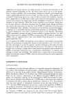
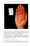
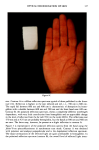
122 JOURNAL OF THE SOCIETY OF COSMETIC CHEMISTS troscopy, Edwards et al. (12) confirmed the earlier observation of Dent (9) that light penetration into whole skin was proportional to wavelength. Jacquez and Kuppenheim (13) confirmed that the erythemal condition was associated with the preferential ab- sorption from a white light source of green light by hemoglobin. However, the demon- stration by Jacquez et al. (14) that electronic measurements of intensity could be made to an absolute accuracy of 1.0%, clearly showed the potential of these devices to mea- sure very small changes in reflected light intensity. In the last two decades a number of small portable devices have been used to measure erythemal values in skin (15,16) with an accuracy and discrimination superior to that of the human eye (17, 18). The remaining problem when considering the evaluation of the clinical condition of skin is the impartiality of the electronic measuring device. Unlike the trained observer, a photosensor has no discretionary powers. Light from the surface of the skin containing topographical information cannot be measured to the total exclusion of light reflected from the bulk tissue containing information on physiological response. It was the aim of this investigation to construct an illumination system for the assessment of the clinical condition of skin which would highlight the surface condition to the exclusion of the erythemal condition or vice versa. To this end a polarized illumination system was developed. EXPERIMENTAL POLARIZED PHOTOGRAPHY Although preliminary photography was successfully developed using a Polaroid SX70 camera to produce instant color prints, it was found that better results were obtained with higher quality photographic equipment as below. The skin area was illuminated from opposite sides at approximately 20 degrees by two Xenon flashlamps (Broncolor Impact) and photographed from 1 m with a Has- selblad camera and 120-mm telephoto lens at f32 using Kodak Vericolor-2 type-S film. At least three photographs were taken of each specimen. A standard photograph for reference purposes was taken. Photographs of reflected polarized and depolarized light from the same specimen illuminated by polarized light were also taken. To achieve this, the system was modified by placing, over each flash lamp, a polaroid filter (18 inches by 18 inches) orientated to make parallel both the polarization vectors of each light beam and also the horizontal specimen plane. A third, rotatable, polaroid filter was fitted to the camera lens. When set parallel to the other polaroid filters, a polarized reflected light image was recorded whilst a perpendicular orientation gave a depolarized reflected light image. In each photographic field a bright aluminium plate partly covered with Kodak white reflectance coating was included as a reference for polarized and depolar- ized light intensity. REFLECTION SPECTROSCOPY Diffuse reflectance spectroscopy was undertaken with a Perkin-Elmer Lambda 7 UV/ VIS spectrometer, at a fixed 4-nm bandpass interfaced to a Perkin-Elmer 3600 UV data station. This standard transmission spectrometer was modified with an external inte-
Purchased for the exclusive use of nofirst nolast (unknown)
From: SCC Media Library & Resource Center (library.scconline.org)