240 JOURNAL OF THE SOCIETY OF COSMETIC CHEMISTS denatured protein. The membrane was then mounted on a Franz diffusion cell and treated with 3 ml of 10 mM calcium chloride solution and dried over a period of 5 days before use in diffusion experiments. Characterization of the model membranes Electron microscopic studies. Scanning electron microscopy was carried out to determine if the membrane surface was uniformly covered with lipid. Model membranes were mounted on an aluminum holder and sputter coated (Sputter Coater, Polaron Instru- ments, Model ES 100) to a 15-mm thickness with gold-platinum alloy prior to scanning (International Scientific Instruments, Model DS 130). Transmission electron microscopy was employed to demonstrate the existence of bilayer structures within the lipid matrix of the model membrane. The specimen preparation involved 30 min hydration of the membrane with buffer 1 hr fixation in 2% glutaraldehyde and 3 hr post-fixation with 1% ruthenium tetroxide (both from Polysciences Inc., Warrington, PA) and serial dehydration with ethanol and final embedment in Spurr's resin (also from Polysciences Inc.). The blocks were then sectioned with a diamond knife and examined by a trans- mission electron microscope (Phillips Electronic Instruments, Model EM-400) operated at 60 KV. Resistance to water transport.' Water vapor transmission experiments across model membranes. Model membranes were sized to 15-mm diameter and mounted on top of a 50-ml glass vial containing saturated MgCO 3 solution yielding --65% relative humidity at 30øC. The vial was then sealed and incubated in a closed chamber at 32øC. Water evaporation through the model membrane was evaluated by periodic determination of the weight loss from the vial over a 24-hr period (13). The weight loss from an aluminum foil- covered vial served as the negative control, and that from an untreated filter served as the positive control. The cumulative weight losses for the three cases were plotted as a function of time. The flux of water across the model membrane was determined after equilibrium was established. Diffusion experiments: Permeability of markers across model membranes. At the end of the 5-day period the membrane was dry and ready for use. The receiver compartment was filled with buffer and maintained at 37øC. Care was exercised to ensure the absence of air bubbles between the underside of the membrane and the receiver solution. The receiver solution was constantly stirred with a small Teflon-covered magnet. The set-up was allowed to stand for 30 min to ensure that no leaks were present. Each diffusion cell was calibrated with distilled water to determine receiver capacity. One ml of an aqueous solution of the marker containing sufficient cold drug was then added to the donor. The concentrations of cold marker in the solutions were: 0.1% for sucrose, and saturated solutions for cortisol, estradiol, and progesterone. A minimum of six membranes was used for each marker, and all experiments were carried out under non-occluded condi- tions. At predetermined time periods, approximately 0.3-0.4 ml of the receiver solu- tion was withdrawn gently using a 1-ml syringe with Teflon tubing attached to the needle via the receiver spout. Before withdrawal of the sample, the solution in the spout was mixed thoroughly with the rest of the solution without the generation of air bubbles or the creation of back-pressure effects. The samples were collected in pre-weighed scintillation vials to accurately determine the weights of solution withdrawn. Fresh buffer, equivalent to that of the withdrawn solution, was then added back to the receiver compartment to maintain constant volume. The samples were then mixed with 10 ml

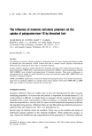
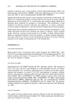
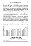
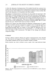
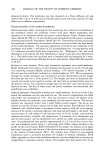
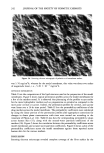
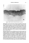
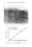
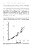
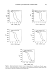
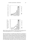
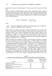
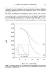
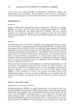
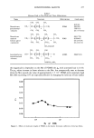
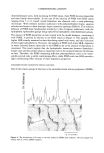
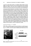
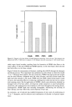
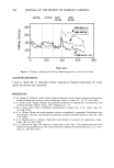
ARTIFICIAL MEMBRANES 241 of Ecolite scintillation cocktail and assayed for radioactivity with a Beckman LS 9000 counter. RESULTS ELECTRON MICROSCOPY Scanning electron microscopy revealed that the model membrane surface was completely covered with a lipid film, although the surface was fairly uneven. Typical micrographs are shown in Figures la and lb. Few lipid crystals could be observed. Figures 2a and 2b show typical transmission electron micrographs of cross-sections of the model mem- branes. Distinctive bilayer patterns are evident at higher magnifications (Figure 2b). The apparent defects seen as empty spaces extending across the sections at low magni- fications (Figure 2a) were found to be artifacts caused by the non-uniform expansion of the bilayers during the fixation process. WATER VAPOR TRANSMISSION STUDIES Figure 3 shows a plot of the cumulative weight loss as a function of time for the model membrane system and for an untreated filter. The water flux across the untreated filter Figure la. Scanning electron micrograph of 2-Ix pore size Nucleopore filter surface. .-
Purchased for the exclusive use of nofirst nolast (unknown)
From: SCC Media Library & Resource Center (library.scconline.org)