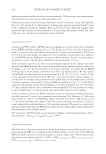
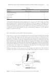
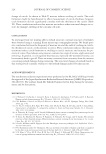
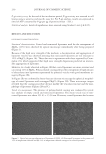
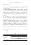
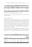
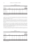
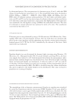
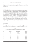
JOURNAL OF COSMETIC SCIENCE 218 The internal structures of the eyelashes were observed using a differential phase-contrast scanning X-ray microscopic computed tomography (CT) system (4). This system enables to observe density distribution of intact samples by measuring the phase gradient of transmitted X-ray probe. MATERIALS AND METHODS EYELASH SAMPLES Eyelash samples were obtained from 36 Japanese women whose use of eye makeup dif- fered. A questionnaire was used to determine their age and whether the women used mascara and/or an eyelash curler, and if so, with what frequency in last two months. The number of women in each age category is shown in Table I, and the frequencies of two eye makeups are shown in Table II. OBSERVATION OF EYELASH STRUCTURE The measurement was performed at the undulator beamline 20XU of SPring-8 (Hyougo, Japan). We employed a differential phase-contrast scanning X-ray microscopic CT system (4), which has been developed to observe the internal structure of human hair sample (5). The differential phase-contrast image representing the variation of refractive index of sample is obtained by measuring the angle of defl ection of the transmitted X-ray beam. The decrement of refractive index from unity is approximately proportional to the elec- tron density. The electron density is generally proportional to mass density. The samples were set in the air. So, the phase-contrast image reconstruction by the CT system corre- sponds to the mass density map of eyelash samples. The sample setup of the scanning X-ray microscopy system is shown in Figure 1. The eyelash sample was positioned such that the eyelash bulb was uppermost, and regions of the eyelash shaft located 2 mm from the eyelash bulb were scanned. The eyelash samples were observed in the atmosphere. More details of the experimental setup are described elsewhere (4). A focused 8 keV X-ray beam with a diameter of 100 nm was used as a scan probe. The transversal scan pitch was 100 nm. For a CT measurement, transversal scan was repeated Table I Age Distribution of Volunteers Age (years) Number of volunteers 20–30 7 30–40 8 40–50 8 50–60 8 60–70 5

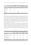
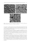
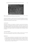
219 INTERNAL STRUCTURE CHANGES OF EYELASH INDUCED BY EYE MAKEUP 600 times by changing the rotation angle by 0.3°—step to cover 180°. In the case of the typical measurement condition with the diameter of the hair sample of 100 μm, more than 1000 points are required for each transversal scan with 100 nm pitch. Therefore, totally more than 600,000 scan points (1000 transversal scan × 600 rotational scan) are required for one CT measurement. In such a measurement condition, it takes approxi- mately 30 min for a CT scan. STRUCTURE ANALYSIS OF RECONSTRUCTED EYELASH IMAGE Shape and size of eyelash. The reconstructed cross-sectional image of eyelash was assumed to be an ellipse. The longest diameter of the cross-sectional image was deemed to be the major axis of the fi ber. The minor axis of the fi ber was assumed the longest diameter per- pendicular to the line of major axis. From the length of major axis and minor axis, the area of the fi ber was calculated. The medulla was assumed to be ellipse- and the length of major axis and that of minor axis were measured by the same method of eyelash fi ber measurement. Thickness of cuticle was estimated by defi ning the average of four locations of cuticle space, two of them were contained in the major axis in the fi ber and the other two were contained in the minor axis of the fi ber. Ranking of the degree of crack formation in cuticle. Cracks were observed in the cuticles of eyelashes. The degree of crack formation was ranked into one of fi ve categories: (i) almost Table II Frequency of Eye Makeup Use Frequency Mascara use (n) Eyelash curler use (n) More than 4 times/week 15 16 2 or 3 times/week 3 4 1 time/week 2 3 Almost never 8 4 Never 8 9 Figure 1. Set up of the scanning X-ray microscopy system with an eyelash sample.
Purchased for the exclusive use of nofirst nolast (unknown)
From: SCC Media Library & Resource Center (library.scconline.org)