JOURNAL OF COSMETIC SCIENCE 18 of the WHO. Several studies have shown that air pollutants interfere signifi cantly with the normal functions of lipids, deoxyribonucleic acid, and proteins in human skin, which is the outermost line of defense of the human body, via oxidative damage (2–10). Those effects lead to skin aging infl ammatory or allergic conditions, such as atopic dermatitis, psoriasis, and acne and skin cancer (11,12). It is easily imagined that the route of air pollutants that damage human health is through inhalation. However, re- cent epidemiological studies regarding the skin have reported that human beings liv- ing in places where they are exposed to much higher levels of air pollutants, such as China and India, suffer from skin problems such as pigmented spots and wrinkles at a high frequency (13). In addition, the number of people who suffer from sensitive skin in Japan increases every year, and about 30% of them feel that the causative factor of their sensitive skin is air pollution (14). In general, it has been demonstrated that PAHs induce infl ammation by activating the aryl hydrocarbon receptor (AhR) signaling pathway (15). AhR is a chemical receptor that responds to exogenous and endogenous chemicals by inducing or repressing the expression of several genes that have protec- tive or toxic effects (15). In that regard, it has been clarifi ed that the activation of AhR induces the overexpression of cytochrome P4501A1 (CYP1A1) and generates excess reactive oxygen species (ROS) (15–17). In daily life, we are frequently exposed to tobacco smoke and exhaust gas, which are types of air pollutants. This raises the possibility that we suffer from adverse effects of aldehyde compounds (ACs), benzo[a]pyrene (BaP), and other chemicals because they are major components in tobacco smoke and exhaust gas. Among various tissues of the body, it is of greatest concern that the skin is intensively infl uenced by air pollutants because of its localization at the outermost surface of the body. ACs cause protein carbonylation in the skin, and it has also been reported that ROS are generated from carbonylated proteins (CPs) when irradiated with blue light through a type-I photosensitization reaction (18). The sum of these facts suggests that ACs initiate an oxidation loop due to the accumula- tion of CPs in the skin. On the other hand, BaP, which is a representative PAH that is produced in industrial combustion processes, enhances the secretion of infl ammatory cyto- kines from human epidermal keratinocytes because of the excess production of ROS through the activation of AhR signaling (19). Thus, it is considered a possibility that air pollutants directly cause these adverse actions because of their penetration through the skin. To avoid that risk, it is very important to develop an effective approach to prevent/ reduce the penetration of air pollutants, PAHs, and ACs, into the skin. Thus, we designed an approach to shield the skin surface using a topically applied fi lm to interfere with the penetration of air pollutants. Sacran, a large molecular weight polysaccharide isolated from Aphanothece sacrum (Suizenji-nori) algae, has unique characteristics and is composed of 11 kinds of monosaccharides, including ionized groups containing sulfate groups and carboxylic acid groups (20–22). We have previously reported that sacran forms a hydrophobic gel-like fi lm in combination with polyols and improves skin conditions in individuals who suffer from atopic dermatitis (23–25). From those results, we expected that sacran would protect the skin against environmental stimuli such as chemicals and air pollutants (e.g., tobacco smoke) because of its formation of an artifi cial skin barrier. Thus, the purpose of this study was to clarify whether sacran reduces skin damage caused by air pollutants focusing on oxidative stress compared with hyaluronic acid (HA), which is commonly used in skin care products.

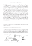
SACRAN PROTECTS SKIN AGAINST POLLUTANTS 19 MATERIALS AND METHODS MATERIALS Sacran was extracted from the algae Aphanothece sacrum (Suizenji-nori) and was purifi ed (19). Bio-sodium hyaluronate (MMW), as a general HA, was obtained from SK Bioland (Chungnam, Cheonan, South Korea), and 1,3-butylene glycol was obtained from Kokyu Alcohol Kogyo Co. (Tokyo, Japan). Dulbecco’s modifi ed Eagle medium (DMEM) and Hanks’ Balanced Salt solution with Ca2+ and Mg2+ (HBSS) were obtained from Nissui Pharmacy (Tokyo, Japan), and fetal bovine serum (FBS) was obtained from Invitrogen (Carlsbad, CA). The BCA Protein Assay Reagent kit was purchased from Pierce Chemical Co. (Rockford, IL). LabCyte EPI-MODEL reconstructed human epidermal equivalents (RHEEs) at 12 d, and their assay medium were obtained from Japan Tissue Engineering (Aichi, Japan). 6-[6-(Biotinylamino)hexanoylamino]hexanoylhydrazine (biotin-AC5- hydrazide) and Hoechst 33342 solution were obtained from Dojindo Laboratories (Kumamoto, Japan). DyLight650-labeled streptavidin was obtained from Thermo Fisher Scientifi c Inc. (Waltham, MA). Acrolein monomer, NBD-hydrazine (4-hydrazino-7-nitrobenzofurazan- hydrazine), and 4-(4,6-dimethoxy-1,3,5-triazin-2-yl)-4-methylmorpholinium chloride were purchased from Tokyo Chemical Industry Co. (Tokyo, Japan). Fluorescein- 5-thiosemicarbazide (FTSC), 2′,7′-dichlorodihydrofl uorescein diacetate (H2DCFDA), and BaP were purchased from Sigma-Aldrich (St. Louis, MO). Dimethyl sulfoxide was purchased from Nacalai Tesque (Kyoto, Japan). 2-Mercapto-ethanol (2-ME) and SYBR® Green Real-Time polymerase chain reaction (PCR) Master Mix for real-time PCR analy- sis were obtained from Thermo Fisher Scientifi c Inc. (Kanagawa, Japan). RLT buffer and the RNeasy Mini Kit were purchased from Qiagen (Hilden, Germany). SACRAN AQUEOUS SOLUTION A 0.05% (w/v) sacran aqueous solution was used for experiments as noted in the text. As a representative anionic polysaccharide that is commonly formulated in skin care products, we used a 0.05% (w/v) HA aqueous solution. BIOTIN CONJUGATION TO POLYSACCHARIDES Polysaccharides were reacted with 2 mg/mL biotin-(AC5)2-hydrazide in the presence of 20 mg/mL 4-(4,6-dimethoxy-1,3,5-triazin-2-yl)-4-methylmorpholinium chloride and pyridine for 5 h. Biotin-conjugated polysaccharides were purifi ed by dialysis against de- ionized water. PENETRATION OF POLYSACCHARIDES INTO RHEEs The penetration of biotin-conjugated polysaccharides through RHEEs was measured by quantifi cation of biotin-conjugated polysaccharides in the culture medium using an Enzyme- linked immunosorbent assay (ELISA) and fl uorescence histology. RHEEs were topically treated with biotin-conjugated polysaccharides, and then were cultured for 24 h at 37°C.
Purchased for the exclusive use of nofirst nolast (unknown)
From: SCC Media Library & Resource Center (library.scconline.org)