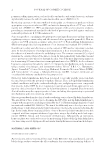
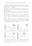
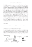
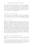
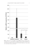
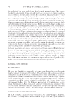
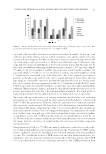
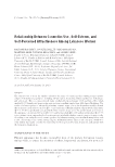
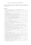
JOURNAL OF COSMETIC SCIENCE 20 Each RHEE was separated into two pieces with a scalpel. Frozen thin sections of one piece of each RHEE embedded in optimal cutting temperature compound were prepared using a cryomicrotome, and the localization of biotin-conjugated polysaccharides in RHEEs was visualized by staining with DyLight650-labeled streptavidin. Nuclei were stained with Hoechst33342. Fluorescence images were taken with a Floid Cell Imaging Station (Thermo Fisher Scientifi c Inc.). The other piece of each RHEE was used to extract biotin-conjugated polysaccharides following homogenization in 500 μL phosphate-buffered saline without Ca2+ and Mg2+ [PBS(-)] at 2,700 rpm for 10 min with a μT-12 bead crusher (Taitec Corp., Saitama, Japan). Biotin-conjugated polysaccharides remaining in the RHEEs or penetrating into the culture medium through the RHEEs were quantifi ed using an ELISA method. Briefl y, the RHEE extract or culture medium was incubated with horseradish peroxidase-conjugated streptavidin (1:1,000) for 1 h at 37°C. The mixed solution was placed in wells of ELISA plates (MS-8596F, Sumitomo Bakelite, Tokyo, Japan) coated with biotin-conjugated bovine serum albmin (BSA), and then incubated for 1 h at 37°C. Each well was washed with PBS-T, then 150 μL 2,2′-azinobis (3-ethylbenzothiazoline- 6-sulfonic acid) diammonium salt (ABTS Wako, Osaka, Japan) solution (0.3 mg/mL) in phosphate-citrate buffer (0.1 M, pH 4.0) containing a small amount of H2O2 was added to each well. After 30 min, the absorbance of each well was measured using a microplate reader (Spark 10M TECAN, Männedorf, Switzerland) at 405 nm. Amounts of biotin- conjugated polysaccharides were determined using a calibration curve prepared with biotin as a standard substance. TOBACCO SMOKE Seven Stars® (tar: 14 mg, nicotine 1.2 mg, Japan Tobacco, Tokyo, Japan) was used a source of tobacco smoke. TRAPPING EFFECTS OF POLYSACCHARIDES AGAINST TOBACCO SMOKE The trapping effects of polysaccharides against tobacco smoke were examined by measuring ACs and BaP in PBS diffused with tobacco smoke using fl uorescence methods (Figure 1). The smoke obtained from burning one piece of tobacco was introduced into PBS stirred with a magnetic stirrer passing through membrane fi lters (10 μm JH, Merck Millipore, Burlington, MA) that had been treated with or without polysaccharide by soaking in 1 mL polysaccharide Figure 1. Method for treatment by tobacco smoke to PBS(-).

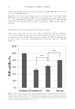
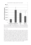
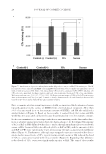
SACRAN PROTECTS SKIN AGAINST POLLUTANTS 21 aqueous solution by aspiration with a vacuum pump, and then dried at 50°C (102 μg/cm2 of polysaccharides were deposited on each fi lter). PBS(-) was reacted with 25 μM NBD-hydra- zine in the presence of 0.05% trifl uoroacetic acid for 30 min in the dark to determine the amount of ACs. ACs were quantifi ed by measuring fl uorescence intensity (FI) (Ex 470 nm, Em 550 nm) using a microplate reader with a calibration curve prepared with acrolein as a standard substance. BaP in the PBS(-) was quantifi ed by measuring FI (Ex 360 nm, Em 450 nm) using a calibration curve prepared with BaP. Trapping effi ciency is expressed as a percent- age versus the value of the nontreated membrane fi lter. CELL CULTURE AND EXPOSURE TO TOBACCO SMOKE HaCaT keratinocytes were cultured in DMEM with 5% FBS at 37°C in a humidifi ed atmosphere containing 5% CO2. HaCaT keratinocytes were inoculated at a density of 3.5 × 10⁴ cells per well in 96-well plates. Cells were cultured in DMEM containing PBS diffused with tobacco smoke in the presence or in the absence of polysaccharides for 24 h. mRNA EXPRESSION OF CYP1A1 The expression level of CYP1A1 mRNA in HaCaT keratinocytes, after culturing for 24 h in DMEM containing PBS diffused with smoke obtained from burning one piece of tobacco by aspiration with a vacuum pump, was evaluated using real-time quantitative PCR analysis. After removal of the medium from wells, washed adherent cells were processed for PCR analyses by the direct addition of 350 μL RLT buffer (Qiagen) containing 3.5 μL 2-ME to each well. Total RNA from the lysed cells was extracted using an RNeasy Mini Kit, according to the manufacturer’s instructions. First-strand cDNA was then synthesized using a PrimeScript RT Master Mix and T100 Thermal cycler (Bio-Rad Laboratories, Hercules, CA). Real-time PCR analysis was performed using an Applied Biosystems StepOne Real-Time PCR System (Thermo Fisher Scientifi c Inc., Kanagawa, Japan) with 1 μL cDNA for each sample. SYBR Green Real-Time PCR Master Mix was used to detect products, and 10 μM concentra- tions of the primers were used: human CYP1A1 and glyceraldehyde-3-phosphate dehy- drogenase (GAPDH) obtained from Takara Bio Inc. (Shiga, Japan) (i.e., NM_002046.5 and NR_045089.1). The relative amount of cDNA in each sample was normalized using GAPDH, and the melting curve was used to verify specifi city. ADVERSE EFFECTS OF TOBACCO SMOKE ON HaCaT KERATINOCYTES The infl uence of smoke obtained from burning one piece of tobacco was measured for the following parameters: cell damage, cell viability, intracellular ROS, and intracellular CPs. Cell damage was examined by measuring cell viability using the neutral red assay. Cells were cultured with DMEM containing 5% FBS and neutral red at a concentration of 33 μg/mL for 2 h. After washing with PBS, neutral red incorporated into the living cells was extracted with 30% MeOH aqueous solution with agitation. The absorbance at 550 nm of the resulting solution was measured using a microplate reader.
Purchased for the exclusive use of nofirst nolast (unknown)
From: SCC Media Library & Resource Center (library.scconline.org)