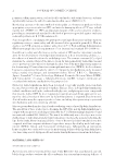
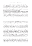
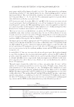
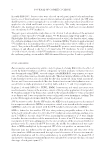
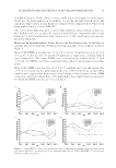
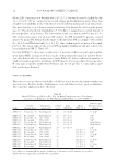
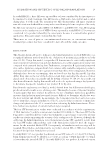
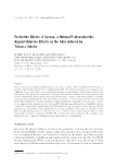
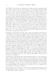
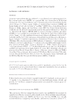
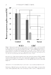
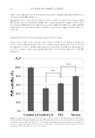
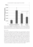
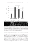
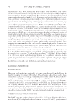
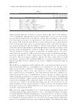
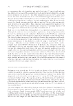
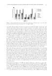
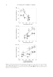
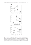
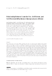
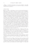
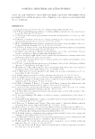
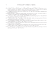
JOURNAL OF COSMETIC SCIENCE 28 First, to examine whether sacran functions as a shield, we monitored the localization of sacran topically applied on the surface of RHEEs from a histochemical viewpoint. More than 99% of sacran stayed in or on the stratum corneum of RHEEs, and HA also exhibited a similar behavior (Figure 2). Based on those results, it seemed likely that sacran and HA would function as an artifi cial barrier because they remained in or on the stratum corneum. In the next examination, to investigate whether sacran remaining on the skin surface func- tions as a barrier against penetration from the chemical aspect, we determined the amount of ACs or BaP in PBS diffused with tobacco smoke through membrane fi lters treated with sacran or HA. Sacran or HA reduced levels of ACs or BaP in PBS, and the amounts of ACs and BaP in PBS were signifi cantly lower after passing through sacran-treated membrane fi lters (Figure 3). Furthermore, although tape-stripped corneocytes treated with tobacco smoke had increased levels of CPs, topical treatment with sacran or HA reduced the level of CPs in corneocytes treated with tobacco smoke (Figure 8). Regarding that ability to reduce levels of CPs, sacran was signifi cantly superior to HA. The sum of these results Figure 7. Amelioration of protein carbonylation induced by tobacco smoke on HaCaT keratinocytes. HaCaT keratinocytes were cultured in DMEM containing PBS diffused with tobacco smoke through fi lters treated with or without sacran or HA. After 24 h, intracellular CP levels were estimated by FI of FTSC labeling. (A) CP levels were quantifi ed by image analysis, and each value represents the mean ± SD of six experiments. Wilcoxon rank-sum test, *p 0.05, ***p 0.001. (B) Representative images of CPs in HaCaT keratinocytes after each treatment (scale bar, 100 μm). Control(-) denotes sham-treated cells and Control(+) denotes cells treated with tobacco smoke diffused through a nontreated fi lter.

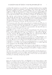
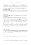
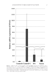
SACRAN PROTECTS SKIN AGAINST POLLUTANTS 29 demonstrates that sacran reduced the penetration of ACs and BaP in tobacco smoke and that the effect was higher than that of HA. To investigate the effects of sacran from the biological viewpoint, we examined the responses of HaCaT keratinocytes to tobacco smoke. In general, it is known that BaP upregulates CYP1A1 through the activation of AhR signaling. CYP1A1 metabolizes PAHs by intro- ducing a hydroxyl group to detoxify BaP. It has been reported that superoxide anion radicals are synthesized as a side product in the metabolic process (15–17). Thus, the ef- fects of sacran as a barrier against tobacco smoke were evaluated regarding CYP1A1 mRNA expression levels in HaCaT keratinocytes. Sacran and HA suppressed mRNA expression levels of CYP1A1 stimulated by tobacco smoke, and, furthermore, the suppres- sion by sacran was signifi cantly higher than that by HA (Figure 4). In addition, sacran ameliorated the cytotoxicity and elevations of intracellular ROS and intracellular CPs Figure 8. Interference with protein carbonylation in corneocytes exposed to tobacco smoke. Tape-stripped corneocytes were treated with sacran or HA, placed in a box fi lled with tobacco smoke for 2 h, and then were incubated for 24 h at 37°C. The levels of CPs in corneocytes was quantifi ed by image analysis. (A) Changes in protein carbonylation in corneocytes. Each value represents the mean ± SD of three independent experiments using the same fi ve volunteers. Wilcoxon rank-sum test, **p 0.01. (B) Representative images of CPs in corneocytes after each treatment (scale bar, 100 μm). Control(-) denotes sham-treated corneocytes and Control(+) denotes corneocytes treated with tobacco smoke without polysaccharide treatment.
Purchased for the exclusive use of nofirst nolast (unknown)
From: SCC Media Library & Resource Center (library.scconline.org)