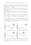
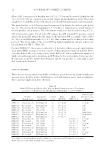
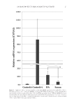
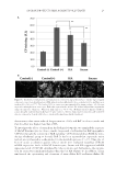
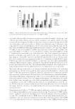
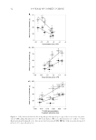
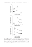
JOURNAL OF COSMETIC SCIENCE 44 more nonionic-based formulations would need to be examined to investigate this theory. The fact that the correlations between CAPB extractions and the skin hydration measures only approached statistical signifi cance is likely because of the small number of formula- tions tested here. A follow-up study to further examine the relationship between CAPB penetration into the skin, SLE1S penetration into the skin, and skin hydration has been completed and will be reported separately. The observation that SLE1S and CAPB have opposite relationships with the clinical mea- sures may be because of differences in these surfactants’ physical properties, and in particu- lar in their structures and/or charges. The structure–function relationships of these surfactants and how they impact the mechanisms of surfactant penetration into human skin are currently unknown. We also noted that the individual surfactants did not penetrate the skin in quantities that are consistent with their bulk solution compositions. For example, the ratio of SLE1S to CAPB in formulation A is 6:1, but CAPB made up 33–39% of the total surfactant mass found in the skin after treatment with formulation A. It is well under- stood that mixed micelle composition often differs from bulk solution composition because of interactions between the surfactants (22). Our results indicate that mixed micelle com- position may play a major role in the mechanism of surfactant-induced skin dryness. More work will need to be carried out in this area to fully understand this phenomenon. CONCLUSIONS Surfactant-induced skin dryness and individual surfactant penetration into human skin were examined using a clinical FCAT study and an ex vivo study using a 2-min exposure protocol. Our results indicate that cup scrub extraction is a suitable substitute for tape stripping in surfactant skin penetration analysis, and may be the preferred method as cup scrubs extract more material and the method is less time-consuming and less expensive to perform. SLE1S and SDS were found to be good predictors of clinical hydration for the anion-based surfactant systems examined here. Furthermore, we found that 14 C-SDS skin penetration from surfactant systems trends with clinical skin hydration induced by the surfactant systems as assessed by corneometry and visual dryness grading, indicating that this ex vivo method may be a useful preclinical test for sulfate-based rinse-off products. More work will need to be completed to understand the structure–function relationships between SLE1S and CAPB and skin penetration, which resulted in oppositely trending relationships for the two surfactants with skin hydration measures. ACKNOWLEDGMENTS Financial support for this research was provided by the Procter & Gamble Company. We thank Chandra Ade-Browne and Tiffany Brooks for their help in conducting the ex vivo skin penetration experiments. REFERENCES (1) P. N. Moore, S. Puvvada, and D. Blankschtein, Challenging the surfactant monomer skin penetration model: penetration of sodium dodecyl sulfate micelles into the epidermis. J. Cosmet. Sci., 54, 29–46 (2003).

SURFACTANT PENETRATION INTO HUMAN SKIN AND RESULTING SKIN DRYNESS 45 (2) A. Lips, K. P. Ananthapadmanabhan, M. Vethamuthu, X. Y. Hua, L. Yang, C. Vincent, N. Deo, and P. Somasundaran, “Role of surfactant micelle charge in protein denaturation and surfactant-induced skin irritation,” in Surfactants in Personal Care Products and Decorative Cosmetics, 3rd Ed. L. D. Rhein, M. Schlossman, A. O’Lenick, and P. Somasundaran. Eds. (CRC Press, Boca Raton, FL, 2006), pp. 177–187. (3) K. P. Ananthapadmanabhan, L. Yang, C. Vincent, L. Tsaur, K. Vetro, V. Foy, S. Zhang, A. Ashkenazi, E. Pashkovski, and V. Subramanian, A novel technology in mild and moisturizing cleansing liquids. Cosmet. Dermatol., 22, 307–316 (2009). (4) V. Goffi n, M. Paye, and G. E. Piérard, Comparison of in vitro predictive tests for irritation induced by anionic surfactants. Contact Dermatitis, 33, 38–41 (1995). ( 5) B. M. Morrison Jr. and M. Paye, A comparison of three in vitro screening tests with an in vivo clinical test to evaluate the irritation potential of antibacterial liquid soaps. J. Soc. Cosmet. Chem., 46, 291–299 (1995). ( 6) M. Almgren, Mixed micelles and other structures in the solubilization of bilayer lipid membranes by surfactants. Biochim. Biophys. Acta Biomembr., 1508, 146–163 (2000). ( 7) C. L. Froebe, F. A. Simion, L. D. Rhein, R. H. Cagan, and A. Kligman, Stratum corneum lipid removal by surfactants: relation to in vivo irritation. Dermatologica, 181, 277–283 (1990). ( 8) T. J. Hall-Manning, G. H. Holland, G. Rennie, P. Revell, J. Hines, M. D. Barratt, and D. A. Basketter, Skin irritation potential of mixed surfactant systems. Food Chem. Toxicol., 36, 233–238 (1998). ( 9) K. D. Ertel, B. H. Keswick, and P. B. Bryant, A forearm controlled application technique for estimating the relative mildness of personal cleansing products. J. Soc. Cosmet. Chem., 46, 67–76 (1995). ( 10) B. H. Keswick, K. D. Ertel, and M. O. Visscher, Comparison of exaggerated and normal use techniques for assessing the mildness of personal cleansers. J. Soc. Cosmet. Chem., 43, 187–193 (1992). ( 11) P. N. Moore, A. Shiloach, S. Puvvada, and D. Blankschtein, Penetration of mixed micelles into the epidermis: effect of mixing sodium dodecyl sulfate with dodecyl hexa(ethylene oxide). J. Cosmet. Sci., 54, 143–159 (2002). ( 12) S. Ghosh and D. Blankschtein, Why is sodium cocoyl isethionate (SCI) mild to the skin barrier? An in vitro investigation based on the relative sizes of the SCI micelles and the skin aqueous pores. J. Cosmet. Sci., 58, 229–244 (2007). ( 13) S. Ghosh and D. Blankschtein, The role of sodium dodecyl sulfate (SDS) micelles in inducing skin bar- rier perturbation in the presence of glycerol. J. Cosmet. Sci., 58, 109–133 (2007). ( 14) M. A. James-Smith, B. Hellner, N. Annunziato, and S. Mitragotri, Effect of surfactant mixtures on skin structure and barrier properties. Ann. Biomed. Eng., 39, 1215–1223 (2010). ( 15) N. McCardy, R. Thompson, M. Miller, P. Styczynski, S. A. Ventura, R. Glenn, and G. B. Kasting, Development of a preclinical surfactant deposition assay to refl ect exposure times typical of consumer use. J. Cosmet. Sci., 68, 219–231 (2016). ( 16) G. B. Kasting, T. G. Filloon, W. R. Francis, and M. P. Meredith, Improving the sensitivity of in vitro skin penetration experiments. Pharm. Res., 11, 1747–1754 (1994). ( 17) R. Kong and R. Bhargava, Characterization of porcine skin as a model for human skin studies using infrared spectroscopic imaging. Analyst, 136, 2359–2366 (2011). ( 18) F. Mohd, H. Todo, M. Yoshimoto, E. Yusuf, and K. Sugibayashi, Contribution of the hair follicular pathway to total skin permeation of topically applied and exposed chemicals. Pharmaceutics, 8, 32–44 (2016). ( 19) S. Grimnes, Pathways of ionic fl ow through human skin in vivo. Acta Derm. Venereol., 64, 93–98 (1984). ( 20) R. R. Burnette and B. Ongpipattanakul, Characterization of the pore transport properties and tissue alteration of excised human skin during iontophoresis. J. Pharm. Sci., 77, 132–137 (1988). ( 21) Y. A. Chizmadzhev, A. V. Indenbom, P. I. Kuzmin, S. V. Galichenko, J. C. Weaver, and R. O. Potts, Electrical properties of skin at moderate voltages: contribution of appendageal macropores. Biophys. J., 74, 843–856 (1998). ( 22) K. Holmberg, B. Jonsson, B. Kronberg, and B. Lindman, “Mixed micelles,” in Surfactants and Polymers in Aqueous Solution, 2nd Ed. (John Wiley & Sons, Ltd., Chichester, West Sussex, England, 2003), 119–138.
Purchased for the exclusive use of nofirst nolast (unknown)
From: SCC Media Library & Resource Center (library.scconline.org)