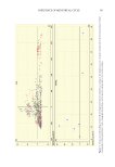
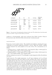
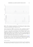
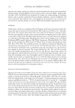
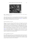
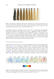
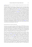
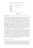
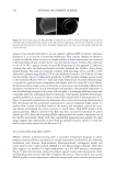
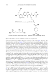
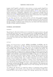
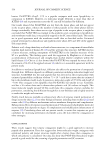
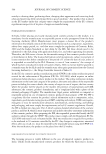
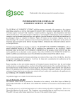
523 CHARACTERIZATION OF BLEACHED HAIR After conditioning for at least 1 h, 3- and 5-µm thick sections were collected in continuous mode using a sectioning speed of 60% maximum. Hair cross-sections were air-dried on paper overnight under reduced pressure. The cross-sections in Figure 1 demonstrate the intrinsic ellipticity of the European dark brown hair fibers. FESEM OF HAIR FIBER CROSS-SECTIONS The cross-sections were imaged using field emission scanning electron microscopy (FESEM) to determine the quality of the sections prior to FTIR imaging and to calculate the cortical cross-sectional area (n = 100). Cross-sections were fixed to aluminum PELCO® pin stubs (Ted Pella, Redding, CA, USA) using 25-mm conductive carbon tabs, and then coated with Au/Pd using our Leica EM ACE600 sputter coater. Finally, the staged fiber cross-sections were imaged with a Hitachi SU-5000 FESEM (Hitachi High Technologies, Schaumburg, IL, USA) using various magnifications. The virgin and bleached European dark brown hair cross-sections were elliptical (see Figure 1), with average major and minor diameters of 80 ± 10 µm and 57 ± 8 µm, respectively. Each fiber contained 5 ± 1 overlapping cuticle cells, and each cuticle cell was 380 ± 90 nm thick. The total pool of analyzed hair fiber cross-sections contained a mixture of medullated and non-medullated fibers. STAGING OF HAIR FIBER CROSS-SECTIONS Prior to FTIR imaging, hair fiber cross-sections must be properly staged on 25-mm calcium fluoride (CaF 2 ) windows (Spectral Systems LLC, Hopewell Junction, NY, USA). A Thermo Fisher Scientific (Waltham, MA, USA) stereo microscope and wooden toothpick were used to move cross-sections into place. In general, approximately 30 cross-sections were carefully juxtaposed without overlap on the CaF 2 crystal surface. Figure 1. FESEM image of European dark brown hair fiber cross-sections (courtesy of W.T. Thompson, Ashland Specialty Ingredients, G.P.).

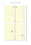
524 JOURNAL OF COSMETIC SCIENCE FTIR IMAGING OF 5-μm HAIR FIBER CROSS-SECTIONS FTIR images were obtained with a Perkin-Elmer Spotlight 400 FTIR imaging microscope (Waltham, MA, USA), which combines an optical microscope with an FTIR spectrometer. The system consists of a linear array of mercury cadmium telluride detectors coupled to a precision automated X-Y sampling stage. Background spectra were collected on sample- free areas of the CaF 2 crystal, and FTIR images were obtained at 8 cm−1 spectral resolution in transmittance mode at 16 scans/pixel. Cross-sectioned hair (5-µm thickness) was used to ensure a linear transmission detector response for the entire IR spectral range. For each of the scans, the spatial resolution of each pixel was 6.25 × 6.25 μm, where each pixel provided a complete mid-IR spectrum. FTIR maps were afterwards generated with ISys software (Malvern Panalytical Ltd., Malvern, UK). The concatenated images were then baseline corrected from the base of the amide I band to 900 cm−1 prior to truncation of the spectra and images (150 × 150 μm). The resulting spectra were processed with GRAMS AI software (Thermo Fisher). FTIR-ATR SPECTROSCOPY OF 3-μm HAIR FIBER CROSS-SECTIONS A Perkin-Elmer Frontier FTIR spectrometer equipped with deuterated-triglycine sulfate detection, and a Perkin-Elmer universal attenuated total reflectance (ATR) accessory was used for all experiments. Small volumes of cryotome-generated 3-μm thick cross-sectioned hair fibers were compressed with a controllable pressure arm against the crystal face of the single-bounce ZnSe ATR crystal. Each spectrum was collected with 64 scans at 4 cm−1 resolution with one level of zero-filling. Perkin-Elmer Spectrum software automatically corrected for water and CO 2 atmospheric contributions, wherein no further vapor correction was needed for subsequent spectral analyses. The resulting spectra were processed with GRAMS AI software. In separate measurements, to evaluate changes in the cuticle composition, complete hair tresses were pressed against the ATR crystal (i.e., cuticles against the crystal). In this case, the goal was to measure oxidative changes to the cuticle of the fiber. Each spectrum was obtained as described previously. RAMAN SPECTROSCOPY OF 3-μm HAIR FIBER CROSS-SECTIONS Raman spectroscopic measurements were performed with a Perkin-Elmer Raman Station 400 F equipped with 785 nm laser excitation. Natural white bleached hair samples that had been micronized to 3-µm sections, as described in the “Hair Cross-Section Preparation” section, were collected, blended, and consolidated for Raman scattering. Blended sections were poured into fresh aluminum DSC pans and loaded into the well plate module for sampling with the Raman laser, which was oriented in the vertical position. Typically, 10 accumulations of 60 sec were performed, with 90 sec exposures occasionally used when photo-bleaching of some background fluorescence was appropriate. As expected in Raman vibrational spectroscopy, the spectral range of the experiment was from 3500–100 cm−1. Spectra were processed with GRAMS AI software.
Purchased for the exclusive use of nofirst nolast (unknown)
From: SCC Media Library & Resource Center (library.scconline.org)