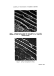
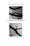
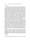
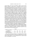
CHEMISTRY OF HUMAN HAIR CUTICLE 359 The morphological progress of digestion in the hair cuticle Examination of digested hair sections in the transmission electron microscope revealed significant progression in the dissolution of the various structural components of the cuticle. In addition, since we believe that the accessibility to enzyme attack of the sectioned hair cuticle and of the physically isolated fragments are probably similar, some correlation could be made between the removal of cuticle components observed in the electron microscope and the various stages of digestion revealed by the foregoing gravimetric results. After digestion of the sections for only 15 min there was almost complete removal of the endocuticle and there was some loss of material from the exocuticle (Fig. 2). (It is pertinent to note that Figs 2 and 3, which are electron micrographs of 'shadowed sections', have been prepared from intermediate negatives so that shadow regions are dark. In achieving this normal consistency for shadows, regions of increasing electron opacity are depicted by increasing brightness in the photographs.) Even at 15 min digestion it is striking that a distinct layer approximately 100 nm wide associated with the cuticle cell boundaries exhibits an electron opacity similar to that of the sectioned epoxy resin, indicating that this component is unaffected by the digestion. After digestion for 45 min virtually all the exocuticle was removed leaving the cell boundary material still apparently unaffected (Fig. 3). At this stage it was necessary to examine metal-stained rather than shadowed sections to reveal the detailed substructure of the cell boundary layer. It was found that this consisted of the A-layer to which was attached, on the outer-facing aspect of the hair, the complete cuticle cell membrane complex (Fig. 4). With further increasing time of digestion the width and general opacity of the A-layer slowly diminished. For periods of digestion exceeding 24 h the A-layer had more or less disappeared still leaving a residue associated with the cell boundaries. Close examination of this revealed that the complete cell membrane complex was still present and bounded on either side by a layer approximately 80 nm thick of stain- able material (Fig. 5). It is noteworthy that at this stage it was difficult to discern the laminated substructure of the membrane complex but this was probably because disorientation of the residue on the electron microscope grid occurs so that the membrane laminae are no longer parallel to the electron beam of the microscope. On the other hand, by examining the point where adjacent cuticle cells overlap to give a type of T-junction of cell boundaries, the initial orientation of the membranes was maintained

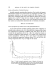
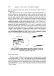
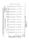
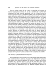
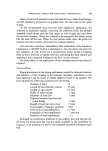
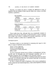
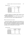
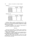
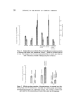
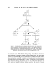
360 JOURNAL OF THE SOCIETY OF COSMETIC CHEMISTS and the laminated substructure of the cell membrane complex could be seen (Fig. 5). From the present results it is clear that the initial fast-dissolving com- ponent observed in the gravimetric studies consists of endocuticle and exocuticle, that the slowly-dissolving component consists of A-layer and that the final insoluble residue consists mainly of cuticle cell membrane material. Indeed there is good correspondence between the percentage areas occupied by the endocuticle + exocuticle + inner layer, the A-layer and cell membrane complex in hair cuticle sections (82-85•o, 10-12•o and 5-6• respectively) and the foregoing gravimetric determinations for what are apparently the same components (82•o, 11•o and 7•o respectively). Little mention has been made of the fate of the cuticle inner-layer in the present work but it is assumed because of its similarity in structure and cystine content (1) that it behaves in a manner analogous to that of the exocuticle. The progress of digestion leading to the isolation in one case of A-layer and cell membrane complex and in the other of the cell membrane complex alone is summarized in the schematic diagram of Fig. 6. Cell membrane corn plex A-layer ..... Exocuhcle Inner layer Endocuticle 90 rnin 72 h Figure 6. Schematic diagram illustrating the course of digestion of human hair cuticle with papain and dithiothreitol and leading to the separation of two major morphological components of the cuticle. Amino acid analysis The first column of Table I contains the amino acid analysis for isolated cuticle and columns 2, 3 and 4, the analyses for the papain/dithiothreitol- insoluble fractions of the cuticle obtained after digestion for 45 min, 90 min and 3 days respectively. The analysis in column 5 was obtained by calculat- ing the differences in amino acid composition between the '90-minute' and '3-day' residues,
Purchased for the exclusive use of nofirst nolast (unknown)
From: SCC Media Library & Resource Center (library.scconline.org)