OESTROGENIC SUBSTANCES IN COSMETICS 309 Test animals Female mice--weight about 20 g--were castrated bilaterally. After 1 week the wounds should have healed and heat phenomena should have disappeared (control with vaginal smear, see below). This was followed by activation with 0.1 ml of 10 [tg oestradiol-1713/ml oil solution subcutaneously on three consecutive days the first sign of oestrogenic action should have appeared about 48 h after the first injection (control by vaginal smear). Animals which did not respond were discarded. This activation was repeated if the animals remained 2 weeks in di- oestrus a lower concentration of oestradiol-1713 (0.5 [tg/ml oil) was used in this case. Reactivation was practised in animals which had not been used for testing during the 2 weeks, as well as in animals which had a negative test result. One week after (re)activation the vaginal smear was checked. Immediately after this check (which should be negative), the animals can be used for the actual test. Mice were discarded if they weighed over 35 g. Administration of samples For screening, the cosmetic product was applied without extraction, on the shaven skin behind the head (the only place that cannot be scratched or licked by the animal). The samples were applied three times, viz. on the first day at 15.00 and on the second day at 10.00 and 15.00. The sample was applied by brush, which was for one sample only. The application was done as reproducibly as possible, and some control of the quantity applied was possible by backweighing. The mice (three per sample) were housed individually in glass jars with a minimum of sawdust. Although the percutaneous test cannot give exact quantitative results, its excellent sensitivity and its simplicity make it the method of choice for screen- ing. Extraction of the sample was only practised when a quantitative test was necessary 1 g of the sample was extracted with 50 ml peroxidefree diethylether. The aqueous layer was discarded. The ether was evaporated and the residue was dissolved in 10 ml 96•o ethanol 0.05, 0.2 and 1.0 ml of this solution were added to 1.5 ml of olive oil. Acetone was added to obtain clear solutions, after which the volatiles were removed under a stream of nitrogen. The remaining oil solution was injected (0.1 ml per animal per injection) at 15.00 (day 1) and at 10.00 and 15.00 (day 2), using 3 animals per concentration. (This was the same scheme as used in the case of percutaneous administration.) Three animals receiving the same treat- ment could be housed in one cage. Preparation and staining of the cytological specimen Vaginal smears were taken by means of a platinum or chromium loop which was adapted to the size of the vaginal opening. The loop was passed through a flame and dipped in distilled water. A droplet of water was placed on an object

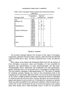
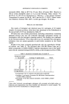
310 JOURNAL OF THE SOCIETY OF COSMETIC CHEMISTS glass with the loop the animal was taken by the tail while gently pressing its back with the third and fourth fingers. The vagina opened itself, so that it was easy to insert the loop. Gently moving the loop back and forth, some vaginal secretion was collected in the loop, which was then dispersed in the droplet on the object glass. One object glass can serve for 9 smears. The preparations were air-dried and fixed for 3 min in methanol. After evaporation of the methanol, the smears were stained in a Giemsa solution diluted 1 : 10, for 20 min, the slides were then rinsed with water and airdried. Microscopical evaluation was done at a medium magnifi- cation without oil immersion. Smears were taken at the following times: Day 3 1500 Day 4 1000 and 1500 Day 5 1000. The smears were evaluated as follows: (a) = (b and c) = (d) = (e and f) = Basic state as seen in juvenile and castrated animals and in mature intact animals in di-oestrus: over two-thirds of the cell material consisted of leucocytes. First sign of beginning of oestrus (comparable with pro-oestrus in intact animals): 1/3-2/3 of the cells were epithelic, either with or without a nucleus. Distinct oestrogenic activity (comparable with beginning of full oestrus in intact animals): over 2/3 of the cells were epithelic, predominantly without a nucleus. Positive (comparable with full heat): over 95•o of the cells were epithelium cells, either with or without a nucleus. The highest score per animal and per group of 3 animals was recorded within one group, reactions should have been comparable if this was not the case, the test should be repeated. If the results of a quantitative test was not 'full heat', but rather stage b, c or d, the test should be repeated with a larger amount of material. Detection limits Some samples of cream-base were tested percutaneously to which known amounts of various oestrogens had been added. The results of these tests are shown in Table I. The limit of detection at percutaneous application lies under 0.00025• for oestradiol-17• and for diethylstilbestrol probably the same is true for oestriol (see Table I). In the quantitative test, the amounts of extract are chosen to obtain responses on the levels of 0.01, 0.025 and 0.0005•o oestrone (or oestradiol-1713- dibenzoate) equivalents per gram. (N.B. 0' 1 pg oestrone or 0.1 pg oestradiol-17[3- dibenzoate = 1 Mouse Unit.)
Purchased for the exclusive use of nofirst nolast (unknown)
From: SCC Media Library & Resource Center (library.scconline.org)