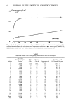
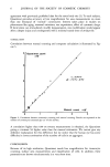
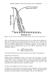
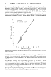
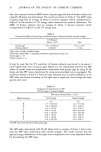
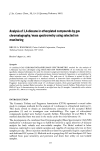
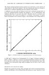
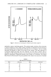
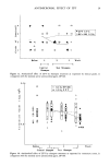
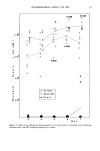
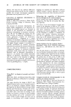
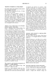
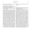
6 JOURNAL OF THE SOCIETY OF COSMETIC CHEMISTS agreement with previously published data for the same body site (3). To each subject, Quantimet provides accuracy at low magnification for area measurements on more than one thousand of "normal" corneocytes. Interest takes place in studies on phenomena like aging, seasonal variations, sun expositions, effect of cytostatic drugs. If local stress can immediately modify desquamation, size modification would appear after a deeper injury and consequently with a minimal transit time of 20 days (9). CORRELATION Correlation between manual counting and computer calculation is illustrated by Fig- ure 5. Quantimet count x10 4 Manual count x 0 • ' • 0 I 2 4 Figure 5. Correlation between automatic counting and manual counting. Results are expressed as the number of corneocytes extracted per cm 2 of skin surface. A correlation higher than 0.99 on twenty measurements is obtained, the Quantimet giving a constant 5% higher value than the manual estimation. We cannot give any definitive explanation for this difference but we dotice that the human eye has some difficulty in correct estimation of the number of cells in an aggregate. CONCLUSION Because of its high resolution, Quantimet needs low magnification for corneocyte counting, surface area measurements and classification of cells. In addition, these parameters are known simultaneously in a very short time.

QUANTITATIVE ASPECTS OF CORNEOCYTES 7 This allows many measurements with good statistical estimation for each subject. The turbine machine is proposed to evaluate natural desquamation by a non-invasive method in man. Fields of application might include cosmetics and dermatology. Then, shape and size measurements of corneocytes during a therapy should yield information about the keratinization process and its modification. ACKNOWLEDGEMENTS We acknowledge the assistance of M. Leveque and the Physics Group who conceived and produced the turbine apparatus. REFERENCES (1) K. J. McGinley, R. R. Marpies and G. Piewig, A method for visualizing and quantitating the desquamating portion of the human stratum corneum,J. Invest. DermatoL, 53, 107-111 (1969). (2) S. Nichols and R. Marks, Novel techniques for the estimation of intracorneal cohesion in vivo, Brit. J. Dermatol., 96, 595-601 (1977). (3) G. Plewig and R. R. Marpies, Regional differences of cell sizes in the human stratum corneum, J. Invest. DermatoL, 54, 13-18 (1970). (4) E. Holzle and G. Piewig, Effects of dermatitis, stripping and steroids on the morphology of corneocytes: A new bioassay,J. Invest. DermatoL, 68, 350-356 (1977). (5) G. L. Grove, Exfoliative cytological procedures as a nonintrusive method for dermatogerontological studies,J. Invest. DermatoL, 73, 67-69 (1969). (6) D. Roberts and R. Marks, The determination of regional and age variations in the rate of desquamation: A comparison of four techniques,J. Invest. DermatoL, 74, 13-16 (1980). (7) M. S. Christensen, S. Nacht, S. L. Kantor and E. H. Gans, A method for measuring desquamation and its use for assessing the effects of some common exfoliants,J. Invest. DermatoL, 71,289-294 (1978). (8) M. F. Stringer and R. R. Marpies, Ultrasonic methods for sampling human skin microorganisms, Brit. J. Dermatol., 94, 551-555 (1976). (9) G. L. Grove, R. M. Lavker, E. Hoelzle and A.M. Kligman, Use of non-intrusive tests to monitor age-associated changes in human skin, J. Soc. Cosmet. Chem., 32, 15-26 (1981). (10) G. L. Grove, R. M. Lavker and A.M. Kligman, Use of microspectrophotometry in dermatological investigations,J. Soc. Cosmet. Chem., 29, 537-544 (1978). (11) H. Goldschmidt, Surface area measurements of psoriatic corneocytes: Effects of intralesional steroid therapy,J. Invest. Dermatol., 73, 558-560 (1979).
Purchased for the exclusive use of nofirst nolast (unknown)
From: SCC Media Library & Resource Center (library.scconline.org)