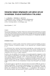
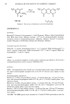
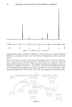
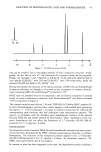
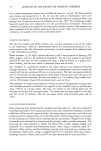
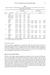
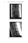
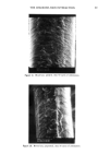
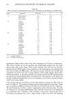
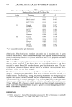
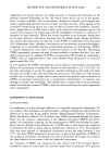
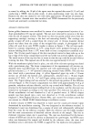
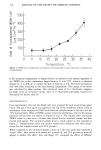
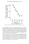
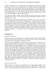
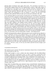
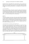
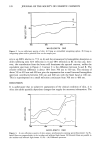
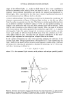
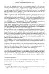
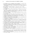
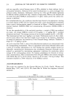
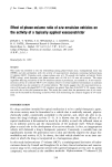
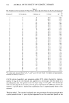
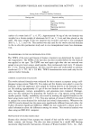
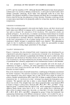
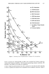
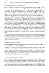
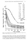
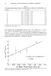
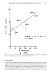
128 JOURNAL OF THE SOCIETY OF COSMETIC CHEMISTS z o too õo B , I • I , 400 500 600 700 WAVELENGTH (NM) Figure 2. In vivo reflectance spectra of skin: A) Using an unmodified integrating sphere. B) Using an integrating sphere with a polaroid filter on the sample port. sity is up (88% relative to 77% in A) and the attenuation by hemoglobin absorption is down (allowing only 32% reflection in A and 48% reflection in B). In this case, how- ever, the contribution from the latter still dominates the spectral contour, unlike the equivalent spectrum in Figure 2. Contour C is the difference between A and B. The relative reflection difference is about 10% from 700 nm to 530 nm. The minima of about 5 % at 620 nm and 600 nm give the impression that a small inverted hemoglobin spectrum contributes between 500 nm and 600 nm with the Soret band at 430 nm. This is superimposed on a small reflection continuum from 700 nm to 400 nm. DISCUSSION It is unfortunate that in subjective assessments of the clinical condition of skin, it is often the subtle spadally dependent changes that supply the necessary information. The •.00. B z 0 50- • m C ' I , I • 400 500 600 700 WAVELENGTH (NH} Figure 3. In vivo reflectance spectra of skin using a synchronously scanning spectrofluorimeter: A) Po- laroid filters are perpendicular in the incident and reflected light beams. B) Polaroid filters are parallel in the incident and reflected light beams. C) (B)--(A) Difference spectrum.

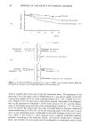
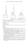
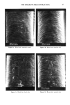
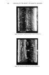
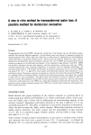
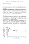
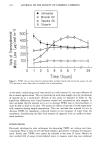
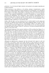
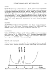
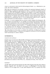
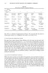
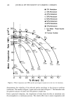
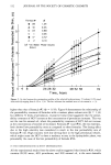
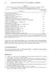
OPTICAL DISCRIMINATION OF SKIN 129 origin of the reflected light, i.e., surface or bulk tissue of skin, is not a problem in subjective assessment since training allows the expert to look at, or into, the skin as required. Using polarized illumination and viewing the polarized and depolarized re- flected light, there is available for the first time a means of preferential selection of pictorial information from different levels in the skin. A clearer understanding of the mechanisms involved can be obtained by considering the schematic representation in Figure 4. Polarized light incident on the skin can reflect from the surface or penetrate into the skin. The former is simply regular reflection from an air-dielectric interface of refractive index 1.5 (19). At near normal incidence no change in the polarization will occur and 100% polarized light will be reflected. There will be some attenuation of the reflected light intensity where the angle of incidence with the non-planar skin surface approaches the polarization angle (20), i.e., in regions showing glare. Light that passes through the air-stratum corneum interface can only return to the surface by reflection from the bulk tissue of the skin. Anderson et al. (21), using diffuse reflection data, concluded that the principal contribution to the remit- tance of light originated in scattering from collagen fibres in the dermis. In the current investigation not only is the scattering changing the direction of the incident light beam through approximately 180 degrees but it is also depolarizing the light. Teale (22) has shown that this is exactly what happens in turbid media, with each scattering event reducing the polarization, expressed as anisotropy, by 0.7 of its orig- inal value. Anisotropy is defined as r = (Ill-IL)/(II1 = 211) where I 11 is the measured light intensity with polarizer and analyser parallel (parallel A B C STRATUH COFLNEUH EP JDE RH ! S DE RH ! S E D Figure 4. Schematic representation of optical paths through skin with polarized light: ¸, Light polarized perpendicular to the plane of the page."O7', Depolarized light. A) Surface reflection. B) Epidermal remit- tance. C) Dermal remittance. D) Forward scattered. E) Dermal scattered. X) Absorption.
Purchased for the exclusive use of nofirst nolast (unknown)
From: SCC Media Library & Resource Center (library.scconline.org)