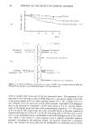
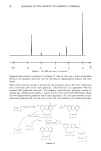
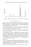
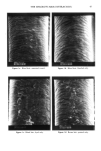
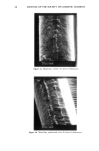
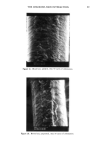
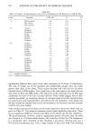
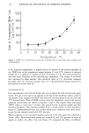
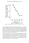
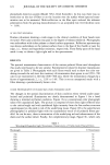
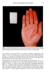
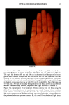
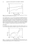
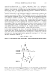
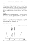
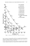
130 JOURNAL OF THE SOCIETY OF COSMETIC CHEMISTS polaroids). I L is the measured light intensity with polarizer and analyser perpendicular (crossed polaroids). Where scattering is most efficient, in the dermis, leading to depolarization, visible light has the highest probability of being absorbed by the hemoglobin in the peripheral blood supply to this region. Each change in direction with a scattering event amounts to an increase in the optical pathlength in an absorbing medium. It is to be expected, therefore, that progressive depolarization would be accompanied by increased spectral attenuation by hemoglobin. As a consequence, there would be a preponderance of re- flected red light and an enhanced contrast of those regions that are normally pink or erythematous. This is exactly the effect demonstrated by the color photography of the depolarized light component of the remitted light. Surface reflected light is all but eliminated by the crossed polaroid on the camera lens since there is a dramatic loss of surface detail when comparing Figure 1C with lB. The enhanced redness in the latter cannot be a photographic artefact or the reference white in each image would show different hues. Such an effect can be seen when comparing the reference whites in Figure lB and 1C with that in 1A where no polaroid filters were present in the optical train. The calculated anisotropy of the remitted radiation at 650 nm (taken from Figure 3), at approximately 0.06, requires multiple scattering to reduce the residual polarization to such a low value. As many as seven scattering events would be required if the anisotropy is reduced to 0.7 for each scattering interaction in the skin. It is not possible to be precise since the calculated anisotropy does not refer uniquely to the dermal remittance but includes polarized reflection from the surface of the stratum corneum. The site-spe- cific sources of the polarized and depolarized light cannot be included in any spectro- photometric corrections that can be undertaken. It must be presumed that the anisot- ropy value is an upper limit. Although the depolarization component has been discussed with respect to only one orientation, the isotropic nature of the light requires an equal intensity for the oppo- site or perpendicular orientation. Both spectral and photographic recordings of the po- larized reflected light therefore contain a depolarized light component. This is consis- tent with the residual erythema shown in Figure lB and the hemoglobin spectrum in Figure 3, contour B. The difference between the polarized and depolarized spectra, contour C, is therefore closer to the corrected polarized reflection spectrum if depolar- ization is complete before remittance of the light from the skin. Although there may be some contribution from an inverted hemoglobin absorption spectrum to the difference spectrum, it is small. No significance can be attached to the absolute intensity, since spectral normalization was undertaken with the polarized and depolarized contours. The featureless nature of the reflection is consistent with the epidermal reflection spectrum published by Anderson et al. (21). The lack of spectral detail in the difference contour gives it a superficial similarity to the polarized reflection spectrum obtained with the integrating sphere, Figure 2, contour B. There is a major inconsistency, however, in that the latter shows much higher absolute reflection intensity, but it must be remem- bered that the difference contour was generated from normalized spectra. Part of the problem relates to the instrumental background correction using a standard white refer- ence overlaid by a sheet of polaroid film. However, application of a sheet of polaroid to the skin with sufficient pressure to ensure a light-tight seal to the integrating sphere would result in compression of the skin. Pressure constriction would reduce blood flow

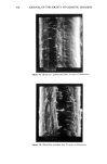
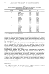
OPTICAL DISCRIMINATION OF SKIN 131 and hence the measured contribution from hemoglobin absorption to the reflection spectrum. Depolarization due to multiple scattering would therefore give higher avail- able light intensities in the absence of hemoglobin, with the parallel oriented vector of the isotropically reflected light contributing to the polarized spectrum. Any additional spectral absorption must be attributed to other light-absorbing moieties that are nor- mally lost under the hemoglobin spectrum. Absence of pressure on the skin is therefore essential if quantitative data are to be compared with the pictorial contrasts shown in Figures lB and 1C. Since no window was present when the spectrofluorimeter was used to record polarized and depolarized spectra, the differences shown in Figure 2 are probably better semiquantitative evalua- tions of the relative pictorial differences of Figures lB and 1C. This result is confirma- tion of the skill of the trained assessor in that all surface topography information is based on evaluating small differences in the amounts of reflected light. When trained assessors score a clinical condition, they can orient the specimen or change the viewing angle in order to optimize the contrast in the feature of interest. The major advantage of polarized and depolarized image recording is in the optimiza- tion of contrast without the need to orient specific regions. Since the polarized image is a composite of polarized and depolarized reflected images, pictorial subtraction of the superimposable depolarized image should leave a difference image of polarized reflected light only. This would have little color and contain only detail of the surface topography of the stratum corneum. If depolarization of light due to surface scattering from rough or dry skin were present, as is suggested by scrutiny of the magnified fine surface detail from Figure 1C, then these features would be atten- uated or lost from the difference image. This may appear to reduce the optical segregating power of the technique but could be used to advantage by pictorial manipulation with an image processor capable of working in color. A measure of the enhanced redness in the depolarized image would allow in vivo quanti- tative analysis to be undertaken with the minimum contribution from skin surface reflection. The latter, however, could still be present as superimposed white or off- white detail since it would arise from fine surface detail depolarization. If discriminating white from pink or red was possible, then the fine surface scattering sites on the skin could be distinguished from the gross surface topography, an operation which the trained assessor does well. The advantage of objective image analysis, how- ever, lies in the very small changes which can be quantitatively determined using an electronic sensor with a finer discrimination of light intensity than is possible by eye. ACKNOWLEDGEMENTS The authors wish to thank Justin North of Graphic Design Section, Unilever Research, Colworth House, for the color photography and preparation of the prints. REFERENCES (1) I. Kanton, W. G. Ballinger, and R. C. Sevin, Severely dry skin: Clinical evaluation of a highly effective therapeutic lotion, Cutis, 30, 410-424 (1982).
Purchased for the exclusive use of nofirst nolast (unknown)
From: SCC Media Library & Resource Center (library.scconline.org)