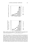
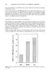
ARTIFICIAL MEMBRANES 239 round-bottomed flask was dissolved in a chloroform-methanol solvent mixture (2:1 by volume). HEPES buffer (0.05 M), pH 7.5, was then added to the organic solution. The ratio of the organic solution to the buffer solution was 2:1 (v/v). The mixture, which was opalescent, became clear after 18 minutes sonication (Branson sonicator, E-module, Shelton, CT). The organic solvents were then removed by a rotary evaporator at 55øC for palmitic acid systems and 70-72øC for the stearic or carnauba acid systems. After solvent removal was complete, the volume of suspension was adjusted with buffer to obtain a suspension containing 30 mg/ml lipid. The liposomes were then annealed for 30 min at the appropriate elevated temperature. The liposomal suspension was then filtered through a 12-p•m pore size Nucleopore TM (Pleasanton, CA) filter to remove any particulate lipids. The filtered suspension was then used immediately to prepare the model membranes. Preparation of membranes Membranes without BSA treatment. One ml of the filtered 30 mg/ml liposomal suspension was diluted threefold with 2 ml of the buffer and extruded through a 2-Ix pore size Nucleopore filter for non-BSA treated membranes and through a 0.45-•m pore size Nylaflo filter (Gelman Sciences, Ann Arbor, MI) for BSA-treated membranes in an extruder (Lipex Biomembranes, Vancouver, BC, Canada). Extrusion was carried out by immersing the entire extruder assembly in a water bath maintained at 40øC, a temper- ature well below the phase-transition temperature of the lipid mixture. The nitrogen pressure used was approximately 40 psi. At the end of the extrusion process, typically an hour, the extruder assembly was dismantled and the lipid-covered nylon filter was carefully retrieved and dried in an oven at 60øC. The dried membrane was then weighed to ascertain the amount of liposomal lipid retained on the filter. For studies of water vapor transmission rates and electron microscopy, the dried mem- brane was treated successively with 2 ml of 3 mM, 5 mM, and 8 mM CaCI 2 solutions in the buffer. Each treatment was separated by 2-hr intervals. The treated membrane was then dried in vacuo for at least 24 hr before use. For diffusion experiments, the lipid filter following extrusion and drying was mounted on a Franz diffusion cell (Crown Glass Co., Somerville, NJ) with a nominal diameter of 2 cm and a receiver capacity of approximately 13 mi. The membrane was placed, lipid side up, on a 0.02-mm thick silastic (Dow Chemical, Midland, MI) cut to O-ring shape, with the inner and outer diameters matching those of the diffusion cell. This was done to ensure a proper seal between the donor cap and the receiver compartment. The donor cap was then placed carefully on the cell and clamped tightly with adjustable clamps. The receiver compartment was left empty and maintained at 37øC. Three ml of 10 mM CaCI 2 solution in pH 7.5 HEPES buffer were then added to the donor and the system was allowed to dry over a period of five days before testing with marker solutions. BSA-treated membranes. The lipid-covered nylon filter, following extrusion and drying, was placed carefully on a glass slide with the lipid side up. Approximately 3 ml of BSA in buffer was then gently added drop-wise to the filter and soaked for 10 min to allow the BSA to spread over the membrane. The filter was then heated at 80øC in an oven for one hour to denature the BSA. This treatment cycle was repeated twice to ensure complete coverage of the filter surface. After the BSA treatment protocol, the surface of the filter membrane exhibited a glossy appearance, indicating the presence of the

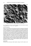
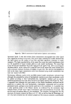
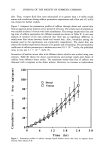
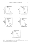
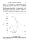
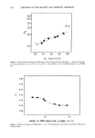
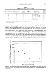
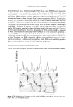
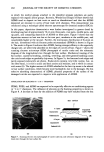
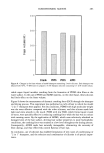
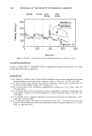
240 JOURNAL OF THE SOCIETY OF COSMETIC CHEMISTS denatured protein. The membrane was then mounted on a Franz diffusion cell and treated with 3 ml of 10 mM calcium chloride solution and dried over a period of 5 days before use in diffusion experiments. Characterization of the model membranes Electron microscopic studies. Scanning electron microscopy was carried out to determine if the membrane surface was uniformly covered with lipid. Model membranes were mounted on an aluminum holder and sputter coated (Sputter Coater, Polaron Instru- ments, Model ES 100) to a 15-mm thickness with gold-platinum alloy prior to scanning (International Scientific Instruments, Model DS 130). Transmission electron microscopy was employed to demonstrate the existence of bilayer structures within the lipid matrix of the model membrane. The specimen preparation involved 30 min hydration of the membrane with buffer 1 hr fixation in 2% glutaraldehyde and 3 hr post-fixation with 1% ruthenium tetroxide (both from Polysciences Inc., Warrington, PA) and serial dehydration with ethanol and final embedment in Spurr's resin (also from Polysciences Inc.). The blocks were then sectioned with a diamond knife and examined by a trans- mission electron microscope (Phillips Electronic Instruments, Model EM-400) operated at 60 KV. Resistance to water transport.' Water vapor transmission experiments across model membranes. Model membranes were sized to 15-mm diameter and mounted on top of a 50-ml glass vial containing saturated MgCO 3 solution yielding --65% relative humidity at 30øC. The vial was then sealed and incubated in a closed chamber at 32øC. Water evaporation through the model membrane was evaluated by periodic determination of the weight loss from the vial over a 24-hr period (13). The weight loss from an aluminum foil- covered vial served as the negative control, and that from an untreated filter served as the positive control. The cumulative weight losses for the three cases were plotted as a function of time. The flux of water across the model membrane was determined after equilibrium was established. Diffusion experiments: Permeability of markers across model membranes. At the end of the 5-day period the membrane was dry and ready for use. The receiver compartment was filled with buffer and maintained at 37øC. Care was exercised to ensure the absence of air bubbles between the underside of the membrane and the receiver solution. The receiver solution was constantly stirred with a small Teflon-covered magnet. The set-up was allowed to stand for 30 min to ensure that no leaks were present. Each diffusion cell was calibrated with distilled water to determine receiver capacity. One ml of an aqueous solution of the marker containing sufficient cold drug was then added to the donor. The concentrations of cold marker in the solutions were: 0.1% for sucrose, and saturated solutions for cortisol, estradiol, and progesterone. A minimum of six membranes was used for each marker, and all experiments were carried out under non-occluded condi- tions. At predetermined time periods, approximately 0.3-0.4 ml of the receiver solu- tion was withdrawn gently using a 1-ml syringe with Teflon tubing attached to the needle via the receiver spout. Before withdrawal of the sample, the solution in the spout was mixed thoroughly with the rest of the solution without the generation of air bubbles or the creation of back-pressure effects. The samples were collected in pre-weighed scintillation vials to accurately determine the weights of solution withdrawn. Fresh buffer, equivalent to that of the withdrawn solution, was then added back to the receiver compartment to maintain constant volume. The samples were then mixed with 10 ml
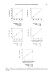
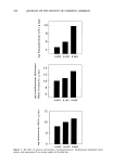
Purchased for the exclusive use of nofirst nolast (unknown)
From: SCC Media Library & Resource Center (library.scconline.org)