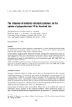
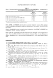
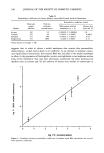
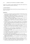
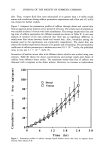
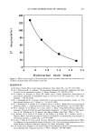
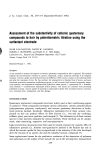
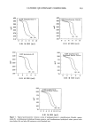
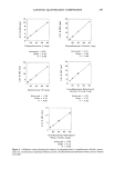
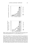
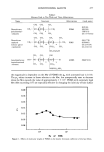
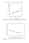
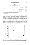
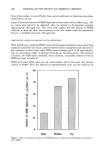
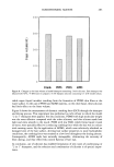
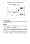
ARTIFICIAL MEMBRANES 247 -11 •, -2 E E -4 -9 -8 -7 -6 -5 -4 -3 -2 log P (human skin) Figure 6. Correlation between permeabilities of markers across BSA membranes and across human skin. stratum corneum with which the lipid bilayer matrix may interact and result in a more cohesive membrane. The BSA-treated membrane, after calcium chloride treatment, exhibited markedly higher resistance to the permeation of sucrose compared to non- treated membranes. It was also confirmed that heat treatment at 80øC of blank filters alone or of lipid membranes without BSA treatment did not lead to reduction of sucrose permeation across these membranes. It was also observed that both BSA and calcium chloride treatment protocols were essential to the generation of model membranes with high barrier properties to hydrophilic drug markers. Treatment of lipid-laden filters with either BSA only or calcium chloride alone did not suffice. This suggests that the model membrane obtained after the combined treatment protocols probably incorporates interactions between the protein and lipid bilayers via calcium-mediated linkages. It would be necessary to carry out extensive structural investigations by electron mi- croscopy and other spectroscopic methods to define the interactions at a molecular level. It is clear, however, that the model membranes prepared in this fashion possess excellent potential to screen a variety of compounds for permeation characteristics across human or animal skin. The excellent correlations of permeability values of several markers across the BSA-model membranes with octanol-water partition coefficients (Fig 5, r 2 = 0.92) and with available data on their permeability across human skin (Fig 6, r 2 = 0.97)

248 JOURNAL OF THE SOCIETY OF COSMETIC CHEMISTS suggest that it is possible to prepare membranes from simple synthetic lipids that are capable of modeling skin permeation characteristics. ACKNOWLEDGMENTS We wish to acknowledge Parrums Christian Dior and Liposome Technology, Inc., for support of this project. REFERENCES (1) D. Kittayanond, C. Ramachandran, and N. Weiner, Development of a model of the lipid constituent phase of the stratum corneum: I. Preparation and characterization of "skin lipid" liposomes using synthetic lipids. J. Soc. Cosmet. Chem., 43, 149-160. (2) C. M. Gary-Bobo, R. Dipolo, and A. K. Solomon, The role of hydrogen bonding in non electrolyte diffusion through dense artificial membranes, Gen. Physiol., 54, 369-382 (1969). (3) R. Dipolo, R. I. Sha'afi, and A. K. Solomon, The transport parameters in a porous cellulose acetate membrane, J. Gen. Physiol., 55, 63-76 (1970). (4) G. L. Flynn and R. W. Smith, Membrane diffusion. III. Influence of solvent composition and permeant solubility on membrane transport, J. Pharm. Sci., 61, 61-66 (1972). (5) F. Bottari, G. DiColo, E. Nannipieri, M. F. Saeton, and M. F. Safarini, Release of drugs from ointment bases. II. In vitro release of benzocaine from suspension type aqueous gels, J. Pharm. Sci., 66, 926-931 (1977). (6) C. R. Behl, E. E. Linn, G. L. Flynn, C. L. Pierson, W. I. Higuchi, and N. F. H. Ho, Permeation of skin and sechar by antiseptics. I: Baseline studies with phenol, J. Pharm. Sci., 72, 391-396 (1983). (7) S. Tanaka, Y. Takashima, H. Murayama, and S. Tsuchiya, Studies on drug release from ointment. IV. Release of hydrocortisone butyrate propionate from topical dosage form to silicone rubber, Int. J. Pharm., 27, 20-38 (1985). (8) W. P. Smith, M. S. Christensen, S. Nacht, and E. H. Gans, Effect oflipids on the reaggregation and permeability of human stratum corneum, J. Invest. Dermatol., 78, 7-11 (1982). (9) W. R. Koch, J. L. Burns, and D. L. Bissett, Preparation and characterization of a reconstituted stratum corneum film as a model membrane for skin transport, Arch. Dermatol. Res., 280, 2152-256 (1988). (10) W. Abraham and D. T. Downing, Preparation of model membranes for skin permeability studies using stratum corneum lipids, J. Invest. Dermatol., 93, 809-813 (1989). (11) S. E. Friberg and I. Kayall, Water evaporation rates from a model of stratum corneum lipids, J. Pharm. Sci., 78, 639-643 (1989). (12) P. W. Hertz and D. T. Downing, Ceramides of pig epidermis: Structure determination,J. LipidRes., 24, 759-765 (1983). (13) G. E. Burch and T. Windsor, Diffusion of water through dead plantar, palmar and dorsal human skin and through toe nails, Arch. Dermatol., 53, 39-41 (1944). (14) G. L. Flynn, S. H. Yalkowsky, and T. J. Roseman, Mass transport phenomena and model: Theo- retical concepts, J. Pharm. Sci., 63, 479-509 (1974). (15) R. J. Scheuplein and I. H. Blank, Permeability of skin, Physiol. Rev., 51, 702-747 (1981). (16) C. Hansch, Ed. Comprehensive Medicinal Chemistry: The Rational Design, Mechanistic Study and Therapeutic Applications of Chemical Compounds, Vol. 6, Cumulative Drug Subject Index 2nd Drug Compendium, Pergamon Press, New York (1990). (17) P. M. Elias, Epidermal lipids: Barrier function and desquamation, J. Invest. Dermatol., 80, 445-495 (1983). (18) P.M. Elias, Epidermal lipids, membranes, and keratinization, Int. J. Dermatol, 20, 1-19 (1981). (19) G. Grubauer, K. R. Feingold, R. M. Harris, and P.M. Elias, Lipid content and lipid type as determinants of the epidermal permeability barrier, J. Lipid Res., 30, 89-96 (1989). (20) P. M. Elias, E. R. Cooper, A. Korc, and B. E. Brown, Percutaneous transport in relation to stratum corneum structure and lipid composition, J. Invest. Dermatol., 76, 297-301 (1981).
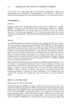
Purchased for the exclusive use of nofirst nolast (unknown)
From: SCC Media Library & Resource Center (library.scconline.org)