JOURNAL OF COSMETIC SCIENCE 66 Wrolstad (23). Anthocyanins were extracted from the lipsticks using equal parts: acetone (70%), acidifi ed ethanol (0.01% HCl), and acidifi ed deionized distilled water, followed by phase partition with chloroform as previously described (21). Sample extracts were diluted in pH 1.0 buffer (KCl-HCl) and in pH 4.5 buffer (sodium acetate) and equili- brated for 15 min. The ACN content, expressed as cyanidin-3-glucoside, was determined using the follow- ing equation: q q1,000 ® pH1.0 pH4.5 Abs Abs DF mg¬ Total monomeric anthocyanin content L ƥqd ( 1) where DF = dilution factor, ε = molar absorptivity of 26,900, and d = path length (1 cm), and the molecular weight of the monomeric ACN content is 449.2. TOTAL PHENOLIC CONTENT The Folin–Ciocalteu method was used to estimate the total phenolic content of the for- mulas containing ACNs, based on the methods described in the literature (24). Lipstick formulas were dissolved in methanol and briefl y sonicated. After sample preparation, they were equilibrated in the dark at room temperature for 2 h before measuring absorbance readings at 765 nm with a spectrophotometer (UV-2450 spectrophotometer Shimadzu). Analyses were conducted in triplicate. Gallic acid was used as a positive control to deter- mine a standard curve for the test. Linear regression was used to determine a standard curve for the absorbance at 765 nm of the gallic acid solutions (R2 = 0.99). The results were reported as mg polyphenolic/L of extract solution as gallic acid equivalents (GAEs). IN VITRO UV ABSORPTION AND SUN PROTECTION FACTOR (SPF) CALCULATION In vitro SPFs for the lipstick formulas were determined based on the methods described by Sayre et al. (25) and Dutra et al. (26) with modifi cations based on the Cosmetics Europe-The Personal Care Association revised method (27). Aliquots of each solution were then pipetted into a UV microwell plate, in eight replications. Absorbance readings were measured by using a SpectraMax190 plate reader (Molecular Devices, Sunnyvale, CA) across the 290- to 400-nm UV wavelength range, at 1-nm increments and blanked against ethanol. The absorbance values were then averaged, and the standard deviations (SDs) were calculated for each sample. The in vitro SPF values were determined according to the following formula: q qI spectrophotometric SPF CF EE AAbs ƫ ƫ (2 ) where CF = correction factor (10), EE (λ) = erythema action spectrum (CIE 1987), I (λ) = spectral irradiance received from the UV source, and Abs (λ) = spectrophotometric absor- bance values at wavelength λ. The EE × I values are constants and were determined by Sayre et al. (25).

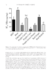
PROPERTIES OF ANTHOCYANIN-PIGMENTED LIPSTICK FORMULATIONS 67 DPPH FREE RADICAL SCAVENGING ASSAY The DPPH free radical scavenging assay was used as a measure of potential antioxidant ca- pacity as described previously (28,29), with modifi cations described by Montanari et al. (11) for use with cosmetics. Methanolic solutions containing the pigments recovered from the lipsticks, the dried pigment extracts at concentrations equal to their contents in the lipstick formulations, and also BHT were used for the testing of antioxidant capacity. DPPH dissolved in methanol with serial dilutions was used to obtain a standard curve for DPPH based on concentration. To the DPPH solution, methanol solutions containing ei- ther BHT, the lipstick base, the dried extract, or the ACN formulas were added in tripli- cate. Samples were equilibrated for 30 min in the dark at 20°C, and absorbance at 515 nm was measured by using a SpectraMax190 plate reader (Molecular Devices) with 30-min equilibration time. Then the samples were blanked against wells containing only methanol. The average of the absorbance readings was then used to determine the DPPH inhibitory percentage and 50% inhibitory concentration (IC50) of each sample. The inhibitory per- centage of each sample was determined based on the following equation: ¬ q100 ® DPPH Sample DPPH Abs Abs % Abs I (3) IC50 values are defi ned as the concentration necessary to inhibit 50% of the free radical (11). The IC50 values of each sample were determined using linear regression of the ab- sorbances at different concentrations. The readings for the ACN formulas were corrected for the absorbance of their respective dried extract in the methanol solution. Samples were tested again after 4 weeks of storage at 4°C, and new IC50 values were calculated and compared with the original values. ANTITYROSINASE ASSAY Tyrosinase inhibition assay was performed as previously reported (20). Methanolic lip- stick extracts and mushroom tyrosinase (5 U) were gently mixed in phosphate buffer (pH 6.5, 50 mM) and incubated for 10 min in a 96-well plate. Absorbance was measured at 475 nm on a SpectraMax190 plate reader (Molecular Devices). Results were compared with the negative control (phosphate buffer) and positive control (kojic acid). The percentage tyrosinase inhibition was calculated as follows: ¬ q100 ® Control Sample Control Abs Abs % Abs I (4) Results are presented as mean (n = 8) ± SD. IC50 values were predicted using linear regression and are expressed in μg/mL. STATISTICAL ANALYSIS Results of the Folin–Ciocalteu method, DPPH free radical scavenging assay, and antity- rosinase activity were analyzed using two-way ANOVA and regression modeling (α = 0.05)
Purchased for the exclusive use of nofirst nolast (unknown)
From: SCC Media Library & Resource Center (library.scconline.org)