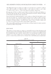
JOURNAL OF COSMETIC SCIENCE 448 benefi ts, as well as in successful cosmetic formulation (4). Most of the investigations on the metabolites derived from seaweed have revealed their potential antioxidant, anti- infl ammatory, anti-wrinkle, anti-bacterial, and anti-aging properties, as well as their role in protection of the skin from ultraviolet rays, moisturizing, and whitening (5). Thus, seaweed is considered as low-costing, safe, and environmentally friendly raw materials for cosmetic industry. Vietnam has a 3,200 km coastline, with great diversity in its algal fl ora. A total of 827 species comprising 88 Cyanophyta, 180 Chlorophyta, 147 Ochrophyta, and 412 Rho- dophyta were compiled from various published sources (6,7). In addition, seaweed farm- ing area in Vietnam has 10.000 ha with the productivity of more than 101.000 tons per year. However, there is no report of the cosmetic applications using Vietnamese seaweed. Until now, researches on seaweed in Vietnam have concentrated on only sampling sur- veys, taxonomy, exploitation of natural bioactive compounds (as fucoidan), and cultiva- tion of economically important seaweed species such as Gracilaria spp., Kappaphycus alvarezii, and K. striatum (8). On the other hand, there still are few studies focusing on nutrition, biochemical composition of seaweed and their applications for functional food, traditional medicines, and biofertilizers (8,9). This study aimed to formulate and evaluate water extracts of four seaweeds, C. lentillifera, S. crassifolium, U. reticulata, and K. alvarezii, in cream mask with anti-aging and moisturizing effects. We fi rst screened potential spe- cies from 10 seaweeds based on the values of some biochemical components as content of polysaccharide, carotenoid, and vitamins. The four selected seaweed species were ex- tracted with water and performed various bioactivities such as antioxidant, antibacterial, cell proliferation, moisture retention, and tyrosinase inhibition in vitro. Finally, formula- tion of cream mask and evaluation of its physiochemical and microbiological characteris- tics from a mixture of four selected seaweeds were investigated. MATERIALS AND METHODS MATERIALS Fresh seaweed species were collected from Nha Trang, Khanh Hoa, Vietnam (12°33′28.9″N 109°17′55.1″E) from February to May, 2017, including Caulerpa lentillifera J. Agardh 1837, Ulva lactuca Linnaeus, 1753, and Ulva reticulata (Forssk) (Chlorophyceae) Kappa- phycus alvarezii (Doty) Doty, and K. striatum (Schmitz) Doty (Pakaya) Gracilaria tenuis- tipitata C. F. Chang & B. M. Xia, 1976 and Gracilariopsis bailiniae J. Zhang & B. M. Xia, 1991 (Rhodophyceae) Sargassum oligocystum Montagne, 1845, S. crassifolium J. Agardh, 1848, and S. denticarpum T. Ajisaka, 1994 (Phaeophyceae). The identifi cation of scientifi c names of these seaweed species was carried out by Tran Mai Duc (Nha Trang Institute of Technology Research and Application, Vietnam Academy of Science and Technology). All seaweed samples were cleaned, rinsed with seawater, dried under dim light, and stored at 2°–4°C until use. METHODS Biochemical composition analysis. The seaweed samples were washed in fresh water and sub- sequently dried at 60°C in an oven the dried samples were ground to particle size 1 mm

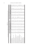
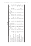
PREPARATION AND EVALUATION OF CREAM MASK 449 and stored at room temperature in airtight plastic containers for biochemical analysis. Protein, lipid, and carbohydrate contents of all seaweed samples were analyzed as previ- ously described (10). The contents of chlorophyll and carotenoid were identifi ed based on the methods by Lichtenthale (11). The vitamin contents were estimated according to the report of Hong and Hien (10). The contents of cadmium (Cd), lead (Pb), arsenic (As), and mercury (Hg) were analyzed by a atomic absorption spectrophotometer (8,10). Preparation of seaweed extracts. 250 g C. lentillifera or 500 g S. crassifolium or 250 g U. reticulata or 100 g K. alvarezii were ground, extracted with 500 mL of distilled water at 60°C for 12 h, and centrifuged at 8,000 rpm/min for 5 min at room temperature. The suspensions were evaporated on a rotary vacuum evaporator to dryness and stored at -20°C until use. Isolation of polysaccharide and fucoxanthin. Carrageenan from K. alvarezii, alginate from S. crassifolium, and ulvan from C. lentillifera and U. reticulata were isolated and determined as described by Aguilana et al. (12) and Cho et al. (13). Fucoxanthin from S. crassifolium was isolated and purifi ed as described by Xia et al. (14). 1-Diphenyl-2-picrylhydrazyl (DPPH) free radical scavenging activity. The antioxidation activity of the seaweed extracts was measured using DPPH radical scavenging assay as described by Harborne and Baxter (15). Ascorbic acid was used as standard control and evaluated for equivalent inhibition (16,17). The inhibition activity of free radicals was calculated in percentage (%) inhibition according to the following formula: % of inhibition = 100 – [(ODs)/(ODc) × 100] with ODs: average optical density of the sample and ODc: average optical density of the control samples (no sample, only DPPH, as 0% inhibitory value). Antibacterial activity. For the antibacterial assay evaluation of seaweed extracts, Escherichia coli ATCC25922, Pseudomonas aeruginosa ATCC27853, Salmonella enterica ATCC13076, Enterococcus faecalis ATCC299212, Staphylococcus aureus ATCC25923, Bacillus cereus ATCC 13245, and Candida albicans ATCC10231 obtained from the National Institute of Food Control, Hanoi, Vietnam, were using as evaluation/testing microorganisms/tools. The bacteria strains were grown on Luria-Bertani nutrient medium (18). Stock solutions of the seaweed extracts, polysaccharide, fucoxanthin, and cream masks were prepared in dimethyl sulfoxide (DMSO), and the antibacterial assays were performed as described previously (19). The minimal inhibitory concentrations (MIC) were recorded as the lowest concentrations inhibiting bacterial and fungal growth. Streptomycin and Cycloheximide were used as positive controls. Mushroom tyrosinase assay. Inhibitory of the seaweed extracts on cell-free mushroom tyrosi- nase activity was determined using spectrophotometry with 3,4-dihydroxyphenylalanine oxidase (DOPA Sigma, St. Louis, MO) as a substrate. Fifty microliter of 0.03% tyrosine solution in distilled water and 75 μL of 0.1 M phosphate buffer (pH 6.8) with the differ- ent concentration of seaweed extracts, polysaccharides, fucoxanthin, and cream masks were added to a 96-well microplate. Finally, 25 μL of mushroom tyrosinase (400 U/mL 0.1 M phosphate buffers) were added, mixed, and incubated at 37°C for 20 min. The amount of DOPA chrome produced in the reaction mixture was determined at 475 nm. Inhibitory effects on the enzyme activity by tested samples were represented as % of in- hibition, [1 - (sample OD475/control OD475)] × 100. Evaluation of moisture retention. The in vitro moisture retention activity of seaweed extracts, polysaccharide, fucoxanthin, and mask creams was assayed gravimetrically as described in a report of Jiménez-Pérez et al. (20).
Purchased for the exclusive use of nofirst nolast (unknown)
From: SCC Media Library & Resource Center (library.scconline.org)