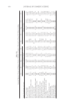
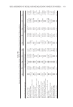
PREPARATION AND EVALUATION OF CREAM MASK 449 and stored at room temperature in airtight plastic containers for biochemical analysis. Protein, lipid, and carbohydrate contents of all seaweed samples were analyzed as previ- ously described (10). The contents of chlorophyll and carotenoid were identifi ed based on the methods by Lichtenthale (11). The vitamin contents were estimated according to the report of Hong and Hien (10). The contents of cadmium (Cd), lead (Pb), arsenic (As), and mercury (Hg) were analyzed by a atomic absorption spectrophotometer (8,10). Preparation of seaweed extracts. 250 g C. lentillifera or 500 g S. crassifolium or 250 g U. reticulata or 100 g K. alvarezii were ground, extracted with 500 mL of distilled water at 60°C for 12 h, and centrifuged at 8,000 rpm/min for 5 min at room temperature. The suspensions were evaporated on a rotary vacuum evaporator to dryness and stored at -20°C until use. Isolation of polysaccharide and fucoxanthin. Carrageenan from K. alvarezii, alginate from S. crassifolium, and ulvan from C. lentillifera and U. reticulata were isolated and determined as described by Aguilana et al. (12) and Cho et al. (13). Fucoxanthin from S. crassifolium was isolated and purifi ed as described by Xia et al. (14). 1-Diphenyl-2-picrylhydrazyl (DPPH) free radical scavenging activity. The antioxidation activity of the seaweed extracts was measured using DPPH radical scavenging assay as described by Harborne and Baxter (15). Ascorbic acid was used as standard control and evaluated for equivalent inhibition (16,17). The inhibition activity of free radicals was calculated in percentage (%) inhibition according to the following formula: % of inhibition = 100 – [(ODs)/(ODc) × 100] with ODs: average optical density of the sample and ODc: average optical density of the control samples (no sample, only DPPH, as 0% inhibitory value). Antibacterial activity. For the antibacterial assay evaluation of seaweed extracts, Escherichia coli ATCC25922, Pseudomonas aeruginosa ATCC27853, Salmonella enterica ATCC13076, Enterococcus faecalis ATCC299212, Staphylococcus aureus ATCC25923, Bacillus cereus ATCC 13245, and Candida albicans ATCC10231 obtained from the National Institute of Food Control, Hanoi, Vietnam, were using as evaluation/testing microorganisms/tools. The bacteria strains were grown on Luria-Bertani nutrient medium (18). Stock solutions of the seaweed extracts, polysaccharide, fucoxanthin, and cream masks were prepared in dimethyl sulfoxide (DMSO), and the antibacterial assays were performed as described previously (19). The minimal inhibitory concentrations (MIC) were recorded as the lowest concentrations inhibiting bacterial and fungal growth. Streptomycin and Cycloheximide were used as positive controls. Mushroom tyrosinase assay. Inhibitory of the seaweed extracts on cell-free mushroom tyrosi- nase activity was determined using spectrophotometry with 3,4-dihydroxyphenylalanine oxidase (DOPA Sigma, St. Louis, MO) as a substrate. Fifty microliter of 0.03% tyrosine solution in distilled water and 75 μL of 0.1 M phosphate buffer (pH 6.8) with the differ- ent concentration of seaweed extracts, polysaccharides, fucoxanthin, and cream masks were added to a 96-well microplate. Finally, 25 μL of mushroom tyrosinase (400 U/mL 0.1 M phosphate buffers) were added, mixed, and incubated at 37°C for 20 min. The amount of DOPA chrome produced in the reaction mixture was determined at 475 nm. Inhibitory effects on the enzyme activity by tested samples were represented as % of in- hibition, [1 - (sample OD475/control OD475)] × 100. Evaluation of moisture retention. The in vitro moisture retention activity of seaweed extracts, polysaccharide, fucoxanthin, and mask creams was assayed gravimetrically as described in a report of Jiménez-Pérez et al. (20).

JOURNAL OF COSMETIC SCIENCE 450 Activity on the proliferation of fi broblasts. Mouse fi broblasts NIH 3T3 were cultured in Dulbecco’s modifi ed Eagle’s medium/Ham’s F-12 nutrient mixture (DMEM/F-12 3:1 by volume, Sigma) supplemented with 10% Fetal Bovine Serum and 1% penicillin/ streptomycin before treatment. All cells were grown in 5% CO2 at 37°C. In the experiment, 2 × 10⁵ cells were added to each well of a 24-well microtiter plate. After addition of sea- weed extracts, polysaccharides, fucoxanthin, and mask creams into each well, the 24-well plate was maintained at 37°C in a CO2 incubator for 2 d. After the cultivation was com- pleted and DMEM removed, 60 mL of 0.5% MTT (3-[4,5-dimethylthiazol-2-yl]-2,5- diphenyl tetrazolium bromide) and 500 mL of fresh DMEM were added to each well. The plate was maintained in a CO2 incubator for 2 h to allow formazan formation. The quan- tity of formazan produced can be regarded as an indicator of cell density or viability. After dissolving the formazan in DMSO, the absorbance at 565 nm was measured with a micro- plate reader (Multiskan Ex, Thermo Electron Co., Vantaa, Finland). The proliferation of fi broblasts was evaluated by comparing the absorbance with that of the untreated control. Cream mask preparation. The formulation of cream mask was composed by demineralized water (49.95–64.95%), Blanose CMC 7HOF (0.5%), emulsifying wax (7%), propylene glycol (5%), Belsil DM 10 (4%), glycerin (1%), talc JA 24R (12%), lunamer 42 (0.5%), and preservative agent PE 9010 (0.05%), and supplemented with 5 mg/mL of mixture of C. lentillifera, S. crassifolium, U. reticulata, and K. alvarezii extracts with ratio of 1:1:1:1 (w:w:w:w). The control cream mask (CCM) was prepared without seaweed extracts. Physiochemical characteristics of the test cream products. The physiochemical characteristics of the test cream products containing 5 mg/mL mixture of seaweed extract were analyzed, including color, coeffi cient of viscosity, pH, refractive index, heavy metals, and microor- ganism. The color, coeffi cient of viscosity, and refractive index of the test cream products were determined according to Vietnam standard 2627:1993, 2642-1993, and 2640:2007, respectively. The pH of the cream masks was measured at 25°C using the Horiba D-71 LAQUAact Portable pH Meter (Horiba Ltd., Kyoto, Japan). Homogeneity, spread ability, and adhesive tests were performed as described by Hanum and Laila (21). The heavy met- als (arsenic, cadmium, chromium, cobalt, lead, mercury, and nickel) were identifi ed as de- scribed by Hepp et al. (22). Microorganisms (such as Staphylococcus aureus, Candida albicans, Pseudomonas aeruginosa, and Escherichia coli) of the test cream products were determined according to Vietnam standard 4884-1:2015 (ISO 4833-1:2013) and TCVN 6972:2001. Irritation test. The irritation test of cream masks was examined by patch test on 20 female volunteers with the age varied from 30 to 40 years. The quantity of test cream applied per test patch was 20 mg. The test articles were dispensed onto 8 mm Finn Chambers® on Scanpor® Tape (Dpro Scientifi c Sdn. Bhd., Petaling Jaya, Malaysia), and the patch was applied to normal skin on the forearm. The patch was removed after 48 h of patch ap- plication. The treatment sites were assessed for the presence of irritation using a 5-point scale 6 h after patch removal. The degree of irritation was evaluated by visual scoring according to the following scale with grading defi ned as follows: 0 = no reaction 0.5 = barely perceptible, very weak spotty erythema 1 = slight erythematic, spotty or diffuse 2 = moderate erythema and 3 = intense erythema, infi ltration, and possible vesicles (23). Skin moisture analysis. The skin moisturizing effects of the cream masks on 20 female vol- unteers (with the age range of 30–40) were measured using a Skin detector SG-5E (Shenzhen, China) according to the protocol of the manufacturer. Before all measurements, each volunteer washed her forearm with a liquid hand wash and then allowed at least
Purchased for the exclusive use of nofirst nolast (unknown)
From: SCC Media Library & Resource Center (library.scconline.org)