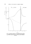
492 JOURNAL OF THE SOCIETY OF COSMETIC CHEMISTS The above hypothesis is supported by the fact that throughout the deficiency-period the water-loss curve and the resistance variation curve have a similar pattern and the decrease in skin resistance corresponds to an increased water-loss. This was to be expected since the rate of water-loss as well as the electrical skin resistance are essentially dependent on the condi- tion of the stratum corneum. The events observed after SSO dosage are indicative of more complex phenomena. Oral ingestion, as expected, causes restoration of the phoreographical response which shows the effect produced on the living cells but, at the same time, there is a considerable increase in resistance and a decrease in water loss. It has been established (5) that any intervention aimed at altering the equilibrium between stratum corneum and germinal cells immediately pro- duces a reaction of the latter in the form of an accelerated keratinization process. After stripping or application of solvent (or detergent solution), skin impedance increases tremendously during the 2 days following the treatment. This is indicative of a keratinization process. Histological studies showed that EFA-deficiency always resulted in a distinct thickening of both the stratum corneum and the malpighian layer (4), also that the mitotic figures were increased. Menton (5) speculated that increased water per- meability of the epidermis might precede or be a stimulus to hyperplasia in an attempt to reduce water-loss by increasing the thickness of the epidermal layer. He observed that EFA-deficient mice kept in a humid atmosphere showed low hyperplasia in the epidermis presumably because of reduced water-loss through the skin. Consequently, it might be thought that for the whole period of time of EFA-deficiency--and owing to the existence of a perturbed stratum cor- neum--considerable mitotic activity is going on. But the division of deficient cells can obviously not restore a normal corneum. This is expressed by an increasing water-loss, decreasing electrical resistance and absence of phoreographical response. With SSO-dosage, however, a normal metabo- lism is restored thereby the germinal cells which had already an increased mitotic rate soon become able to rebuild a normal protective barrier and, thus sheltered, can resume division at the normal rhythm. In other words, the delay observed between the disappearance of the phoreographical response and the increase in water-loss and electrical resistance during the initial stages of EFA-deficiency is due to the existence of a normal mitotic cycle. On the contrary, during the period of restoration following after 7 weeks of EFA-deficiency, a perturbed stratum corneum has been substi- tuted for the former normal one, resulting in an acceleration of the mitotic

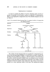
FATTY ACIDS AS REGULATORS OF CELL MEMBRANE FUNCTIONS 493 rhythm and, consequently, reversion to a normal keratinization process a soon as the living cells benefit again from a better balanced lipid metabolism. TENTATIVE INTERPRETATION OF THE PHOREOGRAPHICAL RESPONSE In a previous article (3), we have shown that the variation in skin resistance values which characterizes the phoreographical response origin- ates in the living skin structures. Sufficient experimental evidence is now available which enables us to infer that these living structures are most probably the same as those involved in the psychogalvanic skin reflex, i.e. living epidermal cells and cells of the sweat glands. In the light of the above findings, it seems reasonable to conclude that the phoreographical response is nothing else than the electrical non-linearity common to all biological membranes and which arises from reversible changes in their ion per- meability. It is known that membrane permeation by a given ion species depends on the membrane potential. In the nerve, for example, permea- bility toward sodium ions is governed by the well-known equation of Hodgkin and Huxley (6) and (7) where I m is the flux of sodium ions g the conductance EM, E m the membrane potential at rest and in the excited state respectively. Action potentials of excitable membranes result from the fact that the de- polarization increases gm which, in turn, increases the depolarization. This is why the potential-permeability relationship is not linear. Although in non-excitable membranes the pattern of non-linearity is much less explosive than in excitable membranes, it is necessarily a reflection of the cooperative processes which take place within the membranes. Recent concepts in matter of active transport do relate the phenomena to the existence of molecular carriers which possess several sites for a given ion species. These carriers are seemingly able to assume at least two con- figurational states according to kinetic mechanisms similar to allosteric transitions. The plot of membrane potentials against the rate of transport yields a sigmoid curve.
Purchased for the exclusive use of nofirst nolast (unknown)
From: SCC Media Library & Resource Center (library.scconline.org)