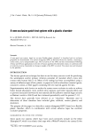
j. Soc. Cosmet. Chem., 34, 1-11 (January/February 1983) The effect of zinc pyrithione on human skin cells in vitro GENJI IMOKAWA and KIKUHIKO OKAMOTO, Kao Soap Company, Ltd., Tochigi Research Laboratories, 2606 Akabane, Ichikai- Machi Haga- Gun, Tochigi,.Japan. Received February 5, 1982. Synopsis A highly active antidandruff agent, zinc pyrithione was investigated on human skin cells (JTC-17) to test the hypothesis that like selenium sulfide, it may also have an antimetabolic action on epidermis, acting on dandruff by reducing increased epidermal turnover. It has been found from the uptake of 3H-thymidine into cells that zinc pyrithione added at levels of 0.25-1.0/xg/ml is effective in suppressing reversibly the DNA synthesis of human skin cells in vitro without a comparable inhibitory effect on RNA and protein synthesis, as shown by the respective uptakes of 3H-uridine and 3H-leucine. Its homologues, sodium pyrithione and omadine disulfide, at levels of 0.25-1.0/xg/ml also exhibit a similar reversible inhibition of DNA synthesis. On long term culture of human skin cells 15-27% growth inhibition was observed with 0.2 /xg/ml of zinc pyrithione. Analysis using synchronized cells revealed that zinc pyrithione can act on all periods of DNA synthesis to suppress it. These findings support the idea that zinc pyrithione may have an antidandruff effect by its antimetabolic action to the skin, rather than its antiyeast action. INTRODUCTION Previous studies to test the relationship between microorganisms and dandruff have revealed that a decreased number of Pityrosporum ovale does not contribute primarily to the reduction in dandruff caused by a highly active antidandruff agent, zinc pyrithione (ZPT) (1). Similar results have been described using selenium sulfide as an antidandruff agent (2). Thus, scalp organisms, especially P. ovale whose increase has been considered as a pathogenic factor for dandruff (3), play no primary important role in production of dandruff. These findings led us to further investigate the possibility that ZPT may have an influence on the scalp skin in a different manner from the way by which ZPT suppresses P. ovale. The amount of dandruff is directly related to cellular reproduction in the basal cells of the skin. In the present study, the effect of ZPT and its derivatives on various cellular metabolisms, especially DNA synthesis, was investigated on cultured skin cells to test the possibility that the reproductive activity by mammalian epithelia might also be suppressed by ZPT.

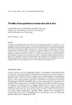
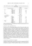
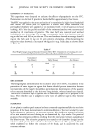
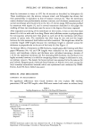
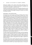
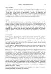
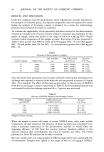
2 .JOURNAL OF THE SOCIETY OF COSMETIC CHEMISTS MATERIALS AND METHODS MATERIALS Eagles Minimal Essential Medium (EMEM) and trypsin (0.25%) were purchased from Grand Island Biological Company. Fetal calf serum (FCS) was obtained from Microbiological Associates. Plastic Petri dishes were purchased from Becton, Dickin- son and Co. 3H-Thymidine (3H-TdR, specific activity 2Ci/mM), 3H-Uridine (3H-UR, specific activity 2-5 Ci/mM), and 3H-Leucine (specific activity 0.5-1 Ci/mM) were obtained from New England Nuclear. Zinc pyrithione (ZPT), Omadine disulfide (DS) and sodium pyrithione (SPT) were obtained from Olin Japan Inc. Irgasan DP-300 (DP-300) was purchased from Ciba Geigy Company. Amethopterin and all other chemicals were purchased from Sigma Chemical Co. CELLS AND CELL CULTURE The JTC-17 human skin cell line (Figure 1), which was derived from a human male bearing an XX sex chromosome constitution (4, 5), was cultured in EMEM Figure 1. Phase contrast microscopy of JTC-17 human skin cells. Note that morphological appearance of the cells has epithelial properties. x 300 supplemented with 10% fetal calf serum, 4 mM glutamine, 100 unit/ml penicillin and 100/•g/ml streptomycin at 37øC with a 5% CO2/95% air atmosphere. Exponentially growing cells were harvested and plated at a cell density of 105 cell/cm 2 in Falcon plastic culture dishes (35mm diameter) containing three 15mm diameter glass coverslips per dish. Medium was replaced three times per week for growth experiments. For the examination of the effect of drugs on the synthesis of DNA, RNA, and protein, 1/•Ci per ml of 3H-thymidine, uridine, and leucine respectively were added to the cultures for a desired period of time. After incubation, cells were washed 3 times with cold Hank's buffer solution. Fixation was done with two successive ten minute treatments with 5%
Purchased for the exclusive use of nofirst nolast (unknown)
From: SCC Media Library & Resource Center (library.scconline.org)