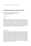
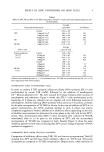
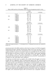
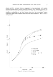
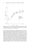
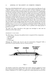
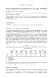
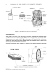
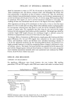
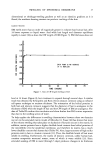
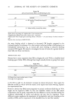
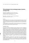
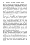
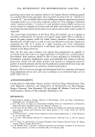
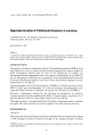
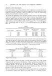
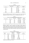
j. Soc. Cosmet. Chem., 34, 21-34 (January/February 1983) Some observations on the swelling of human epidermal membrane C.R. ROBBINS and K.M. FERNEE, Colgate-Palmolive Research Center, 909 River Road, Piscataway, NJ 08854. Received January 27, 1982. Presented at the SCC Annual Scientific Meeting, New York, NY, Dec. 11-12, 1981. Synopsis The swelling behavior of human epidermal membrane has been studied, and results show that the microfibril-matrix models proposed to explain the swelling and extension behavior of wool fiber can also qualitatively account for the swelling behavior of stratum corneum. The outer layers of human skin swell more in thickness than in area, and hair and wool more in diameter than length. This behavior is consistent with the orientation of helical proteins in these three substrates. Epidermal membrane is more sensitive than hair is to water, anionic surfactant, or formic acid, but less sensitive to thioglycolic acid. The greater cystine content of human hair (especially in its matrix) provides a basis for explaining its greater resistance to water, anionic surfactant, and formic acid, and its increased sensitivity to thioglycolic acid. INTRODUCTION The objective of this investigation was to compare the swelling behavior of human epidermal membrane (EM) with other keratins, primarily human hair and wool fiber, to determine if such a comparison might reveal some new structural or functional features about this keratinous substrate. The level of structural organization that controls the swelling behavior of keratin is the secondary and tertiary structure of macromolecular units called microfibrils and matrix (1,2). The microfibrils consist of helical proteins embedded in the less organized matrix, and while in keratin fibers the helical proteins of the microfibrils are oriented parallel to the axis of the fiber (Figure 1) (3), the fibrous proteins of stratum corneum are randomly arranged in planes parallel with the plane of the membrane (Figure 2) (4,5). Thus the diameter of the human hair should be analogous in swelling behavior to that of the thickness of EM, and hair length should be analogous to both the CWL (CW length) and LWL dimensions of EM (Figure 2). These conclusions are confirmed by our experimental findings. 21

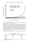
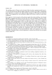
22 JOURNAL OF THE SOCIETY OF COSMETIC CHEMISTS r0•-HELICAL PROTEIN / r PARACORTEX ORTHOCORTEX • / X / F MATRIX f M)CROFIBR IL -- \\ \',.•/f.•\' 11• •-•7 NUCLEAR f •mCROFmBRmL • •00• •0• • •1000 • • aACROFIBRIL CELL {SCALE) Figure 1. Microfibril-matrix structures in wool keratin. EXPERIMENTAL Dark brown "blue string" hair* was used in this study. Diameter was measured before and after treatment on short lengths of fibers (1 to 2 cm) glued to a microscope slide. Measurements were made with a light microscope with attached filar micrometer. Lengths of these same short fiber snippets were also measured using a stereo light microscope with attached filar micrometer. Epidermal membranes were obtained by removal from whole autopsied abdominal STRATUM CORNEUM THICKNESS HAIR •-• CUTICLE j •m CODULLA / MICROFIBRIL ORIENTATION Figure 2. Schematic of human hair and stratum corneum. *De Meo Bros., 135 Fifth Avenue, New York City.
Purchased for the exclusive use of nofirst nolast (unknown)
From: SCC Media Library & Resource Center (library.scconline.org)