228 JOURNAL OF THE SOCIETY OF COSMETIC CHEMISTS quantify the penetration of SLS from aqueous solutions and correlate that to simulta- neous diffusion of water. MATERIALS AND METHODS CHEMICALS Sodium lauryl 35S-sulfate (35S-SLS) and 3H-water were obtained from Amersham, UK. 35S-SLS was purified prior to use to a radiochemical purity 97%, according to a method of Perrin et al. (13). The purity was determined by thin-layer chromatography on silica plates. The plates were developed in isopropanol:chloroform:methanol:water (40:40:20:8 v/v) or in chloroform:ethyl acetate:formic acid (50:40:10). The radioac- tivity on the plates was located using a Berthold LB 2723 radioscanner. Unlabeled SLS was obtained from Henkel Kemi, purity 98%. The donor solution consisted of 35S-SLS and unlabeled SLS dissolved in 3H-water. The SLS concentrations were 0.1, 1.0, and 10% (w/w), with a radiochemical concentration of 1 X 10 •ø Bq/ml. The radiochemical concentration of water was 7 X 10 lø Bq/ml. ANALYSES The radioactivity was measured in a Packard Tri-Carb liquid scintillation counter, model 3375, with Instagel (Packard) as scintillation cocktail. 35S and 3H have different energy levels, thus permitting simultaneous quantitation of SLS and water in the samples by measuring the radioactivity at different energy regions and correcting for quenching and spillover. The radiochemical concentration of the isotopes was chosen so that the number of counts of 35S in the lower energy region was low, compared with the counts due to tritium. PERMEABILITY EXPERIMENTS The penetration experiments were performed according to similar procedures described previously (14, 15,16). Human skin was obtained from cosmetic surgery at the Depart- ment of Plastic Surgery, Sabbatsbergs Hospital, Stockholm, Sweden. Normal skin from three females was used, one abdominal and two breast skin. Due to differences in size, the abdominal skin and one of the breast skin samples were divided into six smaller pieces each, whereas breast skin from the other donor was divided into three pieces. Five experiments were performed on each concentration, and the same combination of skin samples was used (i.e., two abdominal + two breast + one breast skin). After mechanical removal of the subcutaneous fat, pieces of skin were wrapped in aluminium foil and stored at -20øC. Harrison et al. reported that freezing of the skin did not affect the permeability (17). Before starting an experiment, samples were thawed at room temperature. Pieces of skin were mounted in flow-through diffusion cells of stainless steel. The diffu- sion cells were kept in a water bath at 30øC. The SLS solution, 200 Ixl, was applied to the outer surface of the skin. The area available for diffusion was 0.5 cm 2. Beneath the skin, through the lower section of the cell, a receptor medium (normal saline) was pumped and collected in polyethylene vials. The continuous supply (approximately 2

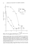
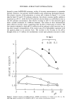
PENETRATION OF WATER AND SLS 229 ml/hr •) of fresh receptor medium assured sink condition throughout the diffusion experiment. The vials were mounted in a fraction collector that changed the position of the vials every 90 minutes. After 49.5 hours the experiments were terminated. CALCULATIONS OF THE RESULTS For each diffusion cell, the cumulative amount of compound penetrating per unit time was plotted against time. Permeability coefficients (Kp, •cm/min l) were determined by dividing the slope of the graph by the concentration of compound applied: J/C = Km D/h = Kp where J is the penetration rate of compound (g/cm2/minl), C is its concentration in the vehicle (g/cm3), Km is the stratum comeurn/vehicle partition coefficient for the drug (without unit), D is its diffusion coefficient in the stratum comeurn (cm2/minl), and h is the thickness of the stratum corneum (cm). RESULTS The time course of the penetration of three concentrations of SLS is shown in Figure 1. 5 • I o E x2 __o 1 E 6 12 18 24. 30 36 42 4.8 TIME (hours) Figure 1. Time course of penetration of 0.1% (A, n = 0) 1.0% ([•, n = 1) and 10.0% (Q, n = 2) SLS. Note the difference in magnification (I0 n) between the curves. Each point represents a mean value + SEM of five determinations (three donors).
Purchased for the exclusive use of nofirst nolast (unknown)
From: SCC Media Library & Resource Center (library.scconline.org)