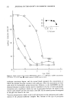
254 JOURNAL OF THE SOCIETY OF COSMETIC CHEMISTS needed for the binding of a reactive hapten to appropriate cells and proteins within skin, and the recruitment and communication with responding T-lymphocytes and other cells. Under physiological conditions, LC continuously present skin-contacting antigens to the T-cells. In this way the skin immune system is continuously made aware of its antigenic microenvironment. Small abrasions and erosions of the skin barrier that nor- mally occur at the microscopic level result in intraepidermal injections of antigenic materials. Other water-soluble compounds will penetrate into the spinal layer of the epidermis and be taken up by LC. When other macrophages fail to clear these antigens, they are processed by the continuously boosted T-cells without any clinical symptoms or signs (2). A large amount of evidence supports the hypothesis that LC are responsible for the critical step of antigen presentation during induction of CH in the skin (13). This evidence includes both circumstantial observations made in vivo and direct observations in experimental animals (3). First, LC are located in relatively large numbers at the cutaneous environmental interface where by implication they should be able to act as APC. Second, experimental work with hapten-derivatized skin grafts has shown that the immunological events that are associated with antigen presentation are accom- plished within the skin graft itself rather than by APC provided by recipients (6). The crucial role of LC in this process is implied by the observation that the major source of IgA immunogenicity in the skin resides with IgA-positive LC (8). It is thus most probably the LC-hapten complex that is involved in presentation of hapten to paracor- tical T-cells. These become activated and are induced to proliferate and differentiate. Thy-1 + DENDRITIC EPIDERMAL CELLS Relatively recently, concepts of cutaneous immunology have been enlarged with the identification in mouse epidermis of a second dendritic cell population, one that ex- presses larger amounts of the cell surface Thy-l-glycoprotein. This cell, termed the Thy-1 + dendritic epidermal cell (Thy-1 + DEC) resembles LC by its marrow derivation (3,9-11), but its expression of Thy-1 antigen suggests an association with T-lympho- cytes or natural killer cells. They resemble primitive T-lymphocytes and may function as epidermal suppressor cells. The functional role of Thy-1 + DEC in humans is un- known, but because it is presumed to be of T-lymphocyte lineage, it is believed to play some role in the cutaneous immune response. KERATINOCYTES, INTERLEUKINS, ETAF Recent work has also demonstrated that the initiation of cellular proliferative responses requires not only physical contact between an antigen-presenting cell (APC) and re- sponding lymphocytes but also the elaboration of a soluble protein, or a "second signal." In the case of T-lymphocyte activation, this second signal is provided by the cytokine interleukin-1 (IL-1), a protein that normally produces the same APC (macro- phage) that is responsible for antigen presentation (12). There is evidence to suggest that epidermal LC produce the monokine IL-1 and present antigens to T-cells. These

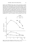
SKIN AND IMMUNITY 2 5 5 data confirm that LC are immune macrophages that provide an immune barrier in the epidermis. While LC have the capacity to produce IL-1, it has also been observed that keratino- cytes have the capacity to produce rather large amounts of a factor that was biologically indistinguishable from IL-1. This factor, termed epidermal cell-derived thymocyte-ac- rivating factor (ETAF), has biologic activities that mimic that of IL-1, indicating that the keratinocyte, believed to be a "non-immune" cell, has the capacity to elaborate at least one immunoregulatory factor, classifying it as an "immunoregulatory cell" (3,6). Not only do keratinocytes produce ETAF, an initiation of T-cell activation, but it has also been proposed that UVR-induced alteration in ETAF production may be respon- sible for several important biological effects of UVR (5). ETAF may even regulate the excessive proliferation of keratinocytes that occurs in psoriasis (14). There is decline of ETAF with age. ETAF represents another mechanism for immune compromise in old skin. The similarity of ETAF to IL-1 is so marked that they are often referred to as ETAF/IL-1 (15). Increase in ETAF occurs when stimulated by an irritant or antigen. Thus, while processing and presenting of an antigen is done principally by LC, they are possibly assisted by the keratinocyte that can elaborate ETAF/IL-1. The neuropeptide alpha-melanocyte-stimulating hormone (ot-MSH) can act as an an- tagonist to IL-1 bioactivities such as the inhibition of thymocyte proliferation. Recently it was reported that topical application of ot-MSH suppresses the cutaneous immune response to contact sensitizers. Further, the loss of contact hypersensitivity (CH) due to applications of ot-MSH could be reconstituted by intradermal or intravenous injections of ETAF/IL-1. Topical application of ot-MSH did not cause alterations of LC. These observations suggest that ot-MSH may represent an additional biologic modifier that can modulate suppression of the CH responses to various haptens (15). This model of epidermal function is made more complex by the observation that factors other than ETAF are also produced by keratinocytes. For example, interleukin-3 (IL-3) is a growth factor that is produced by activated T-lymphocytes and T-cell lines. Both human epidermal cells and squamous cell carcinoma cells have the capacity to produce IL-3. Moreover, there has been derived from epidermal cells a novel factor that inhibits in vitro hypoproliferative responses and IL-2 products (16). This factor, termed epi- dermal cell-derived lymphocyte-differentiating factor (ELDIF) is produced by keratino- cytes. Detailed discussion of the role of interleukins and interferons in immunological skin function is beyond the scope of this review. However, mention should be made that gamma interferon is a lymphokine secreted mainly by activated T-lymphocytes and capable of inducing a wide range of effects on many cells. IL-1 is secreted by macro- phages following uptake of antigen-antibody complexes or following contact with T- lymphocytes during antigen presentation. Secretion of IL-1 is enhanced by gamma in- terferon. IL-1 induces secretion of IL-2, which promotes the growth of T-cells after antigen presentation. IL-6 is a cytokine secreted by phagocytes and activated T-lym- phocytes, as well as by some nonlymphoid cells (e.g., fibroblasts). IL-6 was originally described as a factor that promotes the differentiation of B-lymphocytes to antibody secretory cells.
Purchased for the exclusive use of nofirst nolast (unknown)
From: SCC Media Library & Resource Center (library.scconline.org)