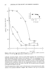
252 JOURNAL OF THE SOCIETY OF COSMETIC CHEMISTS stimulated T-cells in skin release substances that attract macrophages to the site (che- motaxis), prevent them from leaving the site (migration inhibition), enhance their ac- cumulation, and activate them for phagocytosis for increased bactericidal activity and increased secretion of macrophage inflammatory mediators (e.g., prostaglandin pro- teases). The result in a delayed hypersensitivity lesion is a compact focus of highly active cells able to kill the organisms or to render it harmless by sealing off and pre- venting its multiplication and spread (1). Psoriasis has a number of immunopathological cascades contributing to the disease. A major parameter appears to be a gene defect that leads to malfunction of the suppressor T-cells, which prevents recognition of the antigenic epidermal material. The absence of T-cell clones suppresses recognition of basal cell antigens, leading to disturbed matura- tion and impaired keratinization of basal cells, resulting in a self-perpetuating inflam- matory process (1). The predominant defect in the immune system on aging appears to be in the T-cell. In aged persons there is a general depression of the immune response. The number and function of T-cells decrease, and they lose their functional capacities in responding to specific antigens. Age changes in the T-lymphocytes are responsible for much of the pathology that accompanies aging, e.g., cancer. COMPLEMENT, PHAGOCYTES The interaction of antibodies and antigens activates a series of serum proteins collec- tively known as complement. The role of antibodies in complement-mediated effects is to confer specificity on the response. Many of the physiological activities mediated by the humoral immune system are actually carried out by the complement system. The production of antibodies provides a means of identifying foreign molecules within the body. The complement system responds to the signals provided by this recognition system and mediates one or more effector activities, such as phagocytosis, inflamma- tion, cell lysis, and the solubilization of immune complexes. The phagocytic cells of the body are responsible for ingesting and destroying particulate matter. There are two major types of phagocytes: neutrophils and macrophages. Neither of the cells are im- mune cells per se. They are able to engulf foreign particles without any assistance from the immune system. However, the specificity of their action can be greatly enhanced by antibodies. Both neutrophils and macrophages have receptors for a portion of IgG molecules. The phagocytes bind to the antibody-coated targets through these receptors, enhancing phagocytosis. While neutrophils and macrophages share many features, neu- trophils are the most abundant of the circulating white blood cells. Macrophages are localized in such tissues as lymph nodes, spleen, skin, liver, lungs, and wherever for- eign matter gains access to the body. The cell most affected by lymphocyte activity is the macrophage. Lymphocyte products, or direct contact with T-lymphocytes, stimulates macrophages that greatly increase their activity in their allergic reactions. Thus antigen-stimulated T-lympho- cytes, e.g., on the skin, release substances that attract macrophages to the site, prevent them from leaving the site, enhance their accumulation, and activate them for phagocy- tosis. The result in a delayed hypersensitivity lesion is a compact focus of highly active cells able to kill the organism, or render it harmless by sealing it off and preventing its

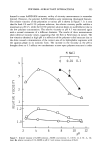
SKIN AND IMMUNITY 253 multiplication and spread (1). In addition to serving as part of the effector arm of humoral immunity, macrophages play an important part in cell-mediated immune re- sponses. The helper T-cell must receive two types of signal in order to respond to a soluble antigen. The T-cell must recognize the antigen in association with a specific macro- phage. In addition, a second signal is non-specific, provided by the cytokine or macro- phage interleukin-1 (IL-1). Thus IL-1 is a lymphocyte-activating factor produced by antigen-presenting cells (APC) in response to antigenic stimuli enhancing T-cell activa- tion (5). LANGERHANS CELLS (LC) The most compelling evidence to support a primary role for skin in immunity concerns observations made about dendritic (branch-like) bone-marrow-derived leucocytes that reside in the epidermis. These are the epidermal Langerhans cells (LC), which are the major source of immunogenicity in the skin. Large numbers of LC form a regular and almost closed network of dendrites within the spinal layer of the epidermis. The large number of LC underlines the antigen-presenting potential of the dendritic cells of the skin. One may hypothesize that LC continuously leave the epidermis, presumably through the lymphatics, being replaced by LC precursors (2). LC comprise about 2% of all epidermal cells. The initial or primary stimulation of an immunological response is induced by an an- tigen, which has been presented by an antigen-presenting cell (APC) or macrophage and responded to by large numbers of lymphatic cells. Lymphold tissues and Langer- hans cells (LC) share macrophage properties and share the roles of fundamental regu- lators of immune function. LC are necessary for initiating T-cell sensitization to an- tigens, as in chronic contact dermatitis. The watershed in concepts concerning the immunological role of skin may be traced to the postulate that skin resembles both gastrointestinal- and bronchial-associated lym- phold tissue by its capacity to attract and retain specific populations of circulatory leukocytes. These cells, which serve specific cutaneous needs, include T-cells and Lang- erhans cells. As an aggregate they were named skin-associated lymphold tissue (SALT) (6). In addition to the SALT category, one may also include a number of immunologi- cally relevant cells that normally populate the epidermal tissues. These include mast cells, tissue macrophages, granulocytes, and veiled cells. All these cells, taken together with those comprising SALT, form an intricate and complex system, which has been called the "skin immune system" (SIS) (2). It has been suggested that SALT represents a physiological mechanism created to deal with neoplastic events taking place contin- uously within the skin due to ultraviolet radiation and the presence of oncogenic viruses and carcinogenic agents (7). LC "recognize" foreign antigens, becoming selective phagocytes taking up particles and substances in solution. Among the substances selectively absorbed are formaldehyde, glutaraldehyde, ethylenediamine, nickel, chromium, and cobalt. LC binds these an- tigens and carries them to the lymph nodes, where lymphocyte activation occurs. They appear to trigger stimulation of T-cells through the antigen. In contact allergic derma- titis (contact hypersensitivity, CH) there is a delayed response reflecting the time
Purchased for the exclusive use of nofirst nolast (unknown)
From: SCC Media Library & Resource Center (library.scconline.org)