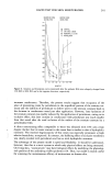
244 JOURNAL OF THE SOCIETY OF COSMETIC CHEMISTS microscopy. Transmission electron microscopy assessments can be applied to biopsy samples, which provide information about the effects of cosmetics on skin (10). How- ever, because of the difficulties encountered in working with biopsies, skin replicas have been obtained by impressing silicone resins, which polymerize easily at ambient tem- perature. This technique has been widely used to determine general skin condition. A negative silicone skin replica is obtained, and from this a polyethylene positive skin replica is developed (10- 13). The positive replicas can be assessed by profilometry (14) or by scanning electron microscopy (15). The aim of our work is to develop a direct method that may provide quantitative information about the efficacy of wrinkle smoothers. The replica technique was used, and further analyses were performed by scanning electron microscopy. The measuring parameters were wrinkle length and width, since their values vary according to the hydrated state of the corneum stratum (16). MATERIALS AND METHODS Two emulsions were prepared: the base formulation (F•) and the same base plus 8% sodium salt of pyrollidone carboxylic acid (Nalidone •-vø) (F2). Both are oil-in-water emulsions. Both formulations were applied in summer to a group of five Caucasian female volun- teers whose ages ranged from 35 to 38 years. They did not suffer from any kind of allergy or cutaneous alteration and were not ingesting any kind of drug. The volunteers had normal skin according to the classification of Koyama et al. (15). A previously selected forearm wrinkle was determined. This comprised an area 1 cm 2, well marked with ink. This area was selected because it was little exposed and had a lower probability of being affected by the environment. Besides, it had low perspira- tion, which permits good skin replicas. All the wrinkles had the same orientation in relation to the arm axis 25 microliters of each formulation were applied over 1 cm 2 of skin. A cross-over design was used, with a 22-day-long non-application period between each treatment of two daily applications during 22 days. At the end of each period, the area under study was evaluated using the selected parameters. Both the wrinkle length, magnified 45 x, and the wrinkle width, magnified 1500 x, were photographed using a JOEL JMS-25 II R scanning electron microscope with a 6 x 7 Mamiya photographic camera adapted to it. A Helios Vernier was used for the determinations. The method used to obtain the replicas was that described by Ryan et al. (12). A Coltene silicone, Coltex Extrafine •!3•, and its catalyst, Coltex Extrafine Mix •!3• were used to obtain the negative skin replica. The positive replica was obtained by using low-den- sity polyethylene (Dow Chemical Co.). RESULTS AND DISCUSSION Before starting the experimental stage, the reproducibility of the technique was checked. For this purpose, two negative replicas of the same wrinkle were taken so as to generate the corresponding positive ones. Microphotographs obtained by scanning elec- tron microscopy were compared as to wrinkle length and width. Starting from one

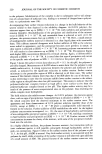
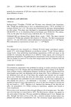
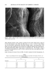
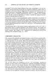
METHOD FOR STUDY OF WRINKLE-REDUCING COSMETICS 245 Figure 1. Scanning electron microscopy (X 45) of a replica utilized to measure a segment of the wrinkle lengths. positive replica, consecutive microphotographs were taken using the same magnifica- tion and compared. This leads to a standardization of the technical conditions for mea- suring. Last, ten consecutive replicas were obtained from one negative replica and the resulting microphotographs were compared. All the determinations showed that there was no difference in length and width of the wrinkle. In order to measure the wrinkle length, a section of the initial wrinkle was selected its ends were clearly defined by the characteristic lines of the skin mosaic. In Figure 1 one can see a microphotograph taken to determine the section of the wrinkle in order to measure the variation of the wrinkle length. On the other hand, in order to measure wrinkle width, the width was considered to comprise the zones of the superposed cor- neous layers of the wrinkle. Figure 2 shows an electron microphotograph taken to determine the wrinkle width, where the superposed corneous layers can be seen. Table I exhibits length determinations in each volunteer after applying the formulations for 22 days. A reduction in wrinkle length can be observed after applying any one of both formulations. This effect is better noticed by calculating the net effect by variation fraction (Table II), where minus sign means diminishing. The length variation fraction is (If - li)/li and the width one is (af - ai)/ai the subindexes i and fstand for initial and final, respectively. Comparing the results obtained when applying formulation F1 during 22 days with
Purchased for the exclusive use of nofirst nolast (unknown)
From: SCC Media Library & Resource Center (library.scconline.org)