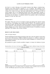
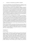
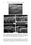
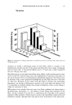
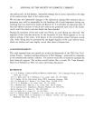
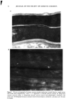
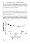
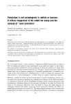
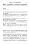
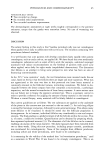
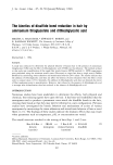
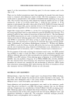
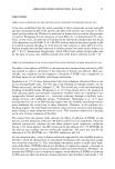
14 JOURNAL OF THE SOCIETY OF COSMETIC CHEMISTS to be no relative displacement of cortical cells. This mechanism of deformation, which is reflected in the stress-strain curve of the fiber, has been discussed extensively in the literature and appears to be well understood and generally accepted. The deformation response of the other major morphological component, the cuticular sheath, is much less well understood. In torsional deformation, Wolfram and Albrecht (6) have found that in the wet state the modulus of the cortex is twenty times greater than that of the cuticula, while in the dry state the torsional properties of cortex and cuticula are similar. In extensional deformation, on the other hand, it is thought that the cuticula does not contribute to the stress-strain curve of the fiber. The cuticle cells are much more rigid in the longitudinal direction and do not have the capacity to release stress by transformation on a molecular level like the cortical cells. Since the cuticular sheath has to deform concomitantly with the deformation of the cortex and the whole fiber, it is assumed that the deformational stress that occurs on the cuticular sheath is released by the movement of cuticle cells relative to each other. The individual cuticle cell is a multilayered structure enveloped by a cellular membrane, the epicuticle, and it adheres to neighboring cuticle cells through the intercellular cement. The outermost cuticle layer is the highly disulfide cross-linked A layer in which 30-35% of the amino acids are half cystine, making it extremely tough and inexten- sible. The next layer, the exocuticle, has a somewhat lower cross-link density and would be expected to be somewhat more easily deformable. The innermost layer is comprised of the much less cross-linked, easily swellable, and highly extensible endocuticle. Dur- ing extension the difference in deformability of the various layers would be expected to result in the generation of shear forces that develop between these layers. At higher extension levels these shear forces can lead to stress concentrations and failure in the weakest layer, the endocuticle. We have studied the response of the surface cuticle of human hair and wool fibers to extension by fluorescence and scanning electron microscopy and will discuss details of that investigation in the following paragraphs. While extension levels used in this investigation are mostly higher than those normally encountered in hair grooming procedures, our results shed light on the deformation processes in the cuticular sheath and its most vulnerable components. EXPERIMENTAL MICROFLUOROMETRY Individual hair fibers are mounted in an extension frame and extended manually under ambient conditions (-50% relative humidity, room temperature) while one observes the fiber in a fluorescence microscope (Leitz MPV 1.1 with Ploemopak attachment). Upon excitation in the ultraviolet range (nonpolarized light, 340-380 nm), the un- treated unextended hair fiber tends to glow with a weak and diffuse blue-white autof- luorescence (Figure 1, left). This intrinsic fluorescence of proteins upon excitation in the UV is based on the presence of certain aromatic amino acids, i.e. tryptophane, tyrosine, and phenylalanine (7,8). It is known that the disulfide groups of cystine are fluorescence quenchers and that their environmental or oxidative scission leads to a significant increase in fluorescence intensity. During extension the surface cuticle edges become

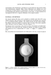
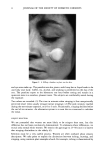
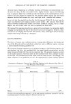
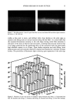
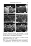
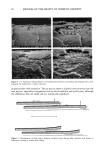
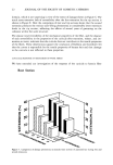
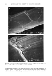
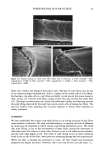
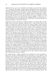
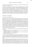
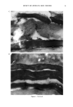
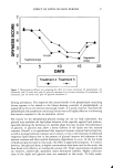
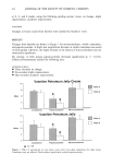
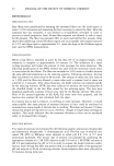
STRESS RELEASE IN HAIR CUTICLE 15 Figure 1. Microfluorometric views of typical hair fiber root sections before (left) and after (right) extension. Excitation at 340-380 nm. visible as they start to move, and brilliant white lines develop at the scale edges as random scale lifting starts in various locations along the hair fiber. It is conceivable that the increased fluorescence intensity at the scale edges during initial fiber extension is indicative of the onset of relative scale movement, revealing endocuticular material that is no longer protected by the quenching effect of the exocuticle with the particularly high disulfide content in its A-layer. Upon further extension and scale lifting, more endocuticular and intercuticular material is exposed and fluorescence intensity increases further. The possibility of fluorescent light scattering upon separation of the scale edge 50 40 -•, lO o Figure 2. Extension of near root section of untreated, brown European hair fibers. A: Unextended hair fiber. B: Random scale lifting. C: Common scale lifting. D: Extreme scale lifting. E: Hair fiber breakage.
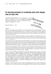
Purchased for the exclusive use of nofirst nolast (unknown)
From: SCC Media Library & Resource Center (library.scconline.org)