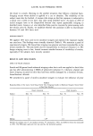
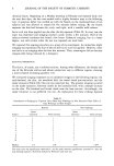
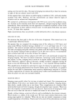
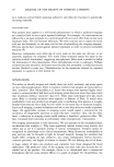
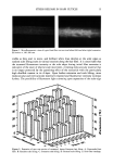
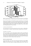
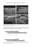
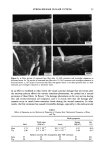
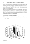
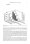
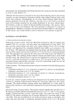
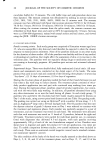
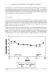
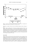
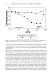
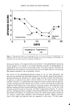
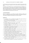
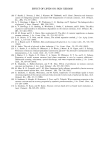
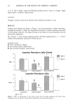
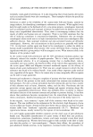
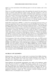
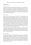
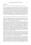
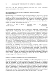
22 JOURNAL OF THE SOCIETY OF COSMETIC CHEMISTS domain, which is not surprising in view of the extent of damage shown in Figure 4. The much more extensive lack of reversibility after the first extension for the tip section is shown in Figure 8. Here the comparison of root and tip sections shows that the second extension produces the various scale-lifting phenomena at considerably lower extension levels in the tip section, reflecting the effects of several years of grooming on the cohesion within the scale structure. The almost total reversibility of the mechanical properties of the fiber, and the absence of such reversibility in the properties of the cuticula after extension, release, and im- mersion in water, indicates that the cuticula does not contribute to the tensile properties of the fibers. These observations support the conclusion of Robbins and Crawford (10) that the cortex is responsible for the tensile properties of human hair and that damage to the cuticula is not reflected in these properties. CUTICULAR RESPONSE TO EXTENSION OF WOOL FIBERS We have extended our investigation of the response of the cuticula to keratin fiber Root Section 40 30 2o lO Figure 7. Comparison of damage phenomena in extended root sections of untreated hair during first and second extensions.

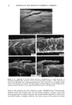
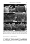
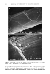
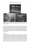
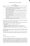
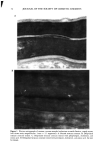
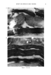
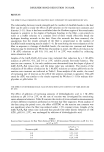
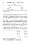
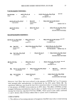
STRESS RELEASE IN HAIR CUTICLE 23 Tip Section 40 30 lO Figure 8. Comparison of damage phenomena in extended tip sections of untreated hair during first and second extensions. extension to include a preliminary study of wool fibers, which in contrast to the multilayered cuticular structure of the human hair fiber show only a single overlapping layer of cuticle cells. A fine pen-fed Merino wool (--25 ptm) and a coarse Coopworth wool (35-40 ptm) were used. Microfluorometry of unextended wool fibers shows diffuse overall autofluorescence when viewed in the UV exitation beam with some highlighting of individual scale edges. In the case of the fine wool, no autofluorescence phenomena suggesting scale lifting could be observed. Only a more pronounced highlighting of scale edges was seen in the form of a fine line. Extension of the coarser wool also failed to produce any scale-lifting phenomena. An increase in fluorescence intensity at the scale edges was observed, but it is impossible to establish whether this increase in brightness is due to scale lifting or simply an indication of stress. SEM studies of the extended fine and coarse wool fibers confirmed the observations in the fluorescence microscope, i.e., the absence of scale lifting upon extension of the fibers. As seen in Figure 9 a,b and 10 a-c, separation at the scale edges is the common response to fiber extension. Failure seems to occur in the intercellular cement of the cell membrane complex, although there may also be some involvement of the endocuticle.
Purchased for the exclusive use of nofirst nolast (unknown)
From: SCC Media Library & Resource Center (library.scconline.org)