28 JOURNAL OF THE SOCIETY OF COSMETIC CHEMISTS influenced by diet, age, race, environment (and seasons), and certain skin conditions (5-7). In psoriasis, for example, stratum corneum cholesterol levels are raised and fatty acids and ceramides decreased, compared with normal subjects (8). The composition of covalently bound lipids in psoriatic stratum corneum also differs from that of healthy stratum comeurn (9). In atopic dermatitis, stratum comeurn ceramide levels, particu- larly ceramide one-linoleate levels, are particularly low (10, 11). Changes in stratum corneum lipid levels are also associated with several hereditary disorders (12), the best-described of which is recessive X-linked ichthyosis (RXLI), in which there is a specific abnormality in sterol metabolism (12), which leads to excessively high levels of cholesterol sulphate in the stratum corneum. Deterioration in skin condition due to seasonal changes in the weather affects far more people than all the above conditions combined, and it is only recently that we have begun to understand the reasons for this situation. Seasonal changes in lipid levels and types occur, with reductions in their levels in winter compared with those in summer (7). Bathing habits also influence the levels of lipids in the superficial layers of the stratum corneum. As a result, in soap-induced winter xerosis, the total levels of stratum corneum ceramides are decreased while those of fatty acids are increased in comparison with normal skin (13-15). The orderly bilayer architecture of these lipids is also dis- rupted in the superficial layers of the stratum corneum (14,15), which probably con- tributes to the increased transepidermal water loss known to occur in these conditions (16). All of these conditions are manifested as dry, flaking skin, which may be more or less severe. The underlying factor that causes the changes in skin structure, function, and appearance in skin xerosis is the failure of the normal desquamatory process, which itself depends upon the specific degradation of intercorneocyte cohesive factors (1,15). For instance, it is known that certain proteases in the stratum corneum are responsible for the normal orderly degradation of desmosomes (17, 18). It has also been reported that enzymic activity, and thereby desmosome degradation, occurs only above a certain water content in the stratum corneum (19,20). When stratum corneum lipid structure is disturbed, the resulting reduction in stratum corneum hydration leads to the retention of corneocytes on the skin's surface and the manifestation of skin xerosis due to reduced desmosome degradation. If, therefore, stratum corneum moisturization and water bar- rier function can be restored by the topical use of suitable lipids and humectants, the desquamatory process may be normalized and xerotic skin conditions may be treated more effectively than they are at present. One compound that is widely recognized for its importance in skin care products is glycerol (21). Products containing glycerol have been shown to be very effective in the treatment of skin xerosis (22), and the action of glycerol has been explained in terms of its occlusive (23), humectant (23,24), and lipid-phase modulating (25,26) properties, all of which translate into moisturization and barrier improvements for the stratum corneum. More recently we have demonstrated that glycerol aids the enzyme lysis of desmosomes in the stratum corneum (19,20). Other occlusive agents also assist to maintain stratum corneum barrier function and improve skin condition. Recently, petrolatum has been shown to penetrate the stratum corneum and interact with stratum corneum lipids (27). Also, stratum corneum ceramides themselves applied topically, particularly in combination with cholesterol and fatty acids, have also been shown to be effective in restoring barrier function in mice with perturbed epidermal barrier function induced by solvents (28), as well as in treating skin xerosis in humans (29,30). The

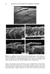
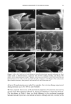
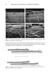
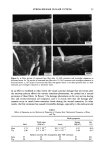
EFFECT OF LIPIDS ON SKIN XEROSIS 29 maintenance of a multilamellar lipid bilayer between the corneocytes has been identified as a key to these benefits (31). Although the skin produces ceramides as the main bilayer-forming lipid in the stratum corneum, we were interested to determine whether other bilayer-forming lipids could mimic their behavior. Phospholipids are the main bilayer-forming lipids found in plasma membranes of living cells. However, they also occur in low levels in the lower layer of the stratum corneum but are usually hydrolyzed in the outer cellular layers. Nevertheless, as they are capable of forming lameliar lipid phases, they may be, there- fore, of use for skin care treatments. We were interested to determine if a mixture of phospholipids, cholesterol, and fatty acids would have beneficial effects in the treatment of skin xerosis, and if these benefits could be enhanced in the presence of glycerol. MATERIALS AND METHODS IN VITRO ELECTRON MICROSCOPY STUDIES Preparation of stratum corneum. Fresh skin (Buckshire Corporation, NJ) was washed with ethanol (70% v/v), cut into 3-cm-wide strips, and dermatomed (0.3-mm thick). The skin was then placed dermis side down onto trypsin solution (0.2% w/v) in sterile, calcium- and magnesium-free, phosphate-buffered saline (PBS), and incubated at 4øC for 18 hours. The epidermis was separated from the dermis and floated epidermis side down on fresh trypsin solution and incubated at 37øC for two hours to release epidermal cells. This procedure was conducted two more times with fresh trypsin and followed by rinsing in PBS. The resulting isolated stratum corneum was floated onto a nylon mesh and desiccated. The isolated stratum corneum was delipidized by extraction in propan- 2-ol (0.1 g SC/100 ml solvent) in a sealed vial at 42øC for one hour and dried. This procedure allowed reproducible extraction of the free intercellular lipids but not the covalently bound stratum corneum lipids. Product treatments. The delipidized stratum corneum was treated with the following three solutions: 1. Chloroform-methanol (2:1 v/v) containing 24 mg/ml of a mixture of phospholipid, cholesterol, and stearic acid (1:2:1 by weight). 2. Chloroform-methanol (2:1 v/v) containing 24 mg/ml of a mixture of petrolatum, cholesterol, and stearic acid (1:2:1 by weight). 3. Chloroform/methanol solution (2:1 v/v). In each case, a known weight of solution was dispensed onto the surface of the stratum comeurn at a loading of approximately 300 I&g/cm 2 and gently rubbed into the stratum corneum. The treated stratum corneum pieces were sandwiched in a nylon mesh to maintain the orientation, and immediately prepared for electron microscopy as described below. Electron microscopy of stratum comeurn. The stratum comeurn samples were pre-fixed in 0.2 M sodium cacodylate-buffered glutaraldehyde solution (2.5% v/v, pH 7.2) for approx- imately 18 hours and then cut into 1-mm-wide strips. The strips were rinsed in cold buffer (0.2 M sodium cacodylate) for 20 minutes followed by a two-hour soak in fresh cold buffer. This step was followed by two further 20-minute rinses in cold buffer. The strips were then post-fixed in ruthenium tetroxide solution (0.2% in 0.2 M sodium
Purchased for the exclusive use of nofirst nolast (unknown)
From: SCC Media Library & Resource Center (library.scconline.org)